- Review
- Open access
- Published:
Spatiotemporal multi-omics: exploring molecular landscapes in aging and regenerative medicine
Military Medical Research volume 11, Article number: 31 (2024)
Abstract
Aging and regeneration represent complex biological phenomena that have long captivated the scientific community. To fully comprehend these processes, it is essential to investigate molecular dynamics through a lens that encompasses both spatial and temporal dimensions. Conventional omics methodologies, such as genomics and transcriptomics, have been instrumental in identifying critical molecular facets of aging and regeneration. However, these methods are somewhat limited, constrained by their spatial resolution and their lack of capacity to dynamically represent tissue alterations. The advent of emerging spatiotemporal multi-omics approaches, encompassing transcriptomics, proteomics, metabolomics, and epigenomics, furnishes comprehensive insights into these intricate molecular dynamics. These sophisticated techniques facilitate accurate delineation of molecular patterns across an array of cells, tissues, and organs, thereby offering an in-depth understanding of the fundamental mechanisms at play. This review meticulously examines the significance of spatiotemporal multi-omics in the realms of aging and regeneration research. It underscores how these methodologies augment our comprehension of molecular dynamics, cellular interactions, and signaling pathways. Initially, the review delineates the foundational principles underpinning these methods, followed by an evaluation of their recent applications within the field. The review ultimately concludes by addressing the prevailing challenges and projecting future advancements in the field. Indubitably, spatiotemporal multi-omics are instrumental in deciphering the complexities inherent in aging and regeneration, thus charting a course toward potential therapeutic innovations.
Background
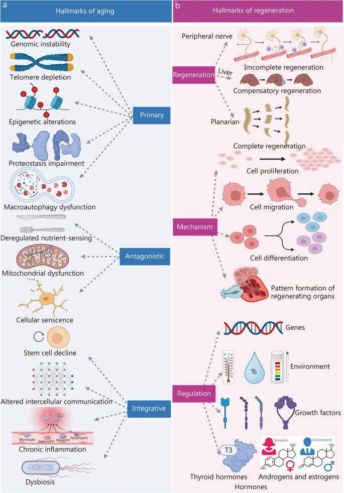
Aging and regeneration, are fundamental biological processes, that have long captivated the scientific community. Aging, a multifaceted phenomenon, is characterized by a progressive decline in physiological functions coupled with an increased vulnerability to age-related diseases [1,2,3,4,5]. To elucidate this complex process, López-Otín et al. [3] introduced a comprehensive framework encompassing twelve hallmarks of aging. These hallmarks, which span a range of molecular, cellular, and organ processes, contribute significantly to aging. They include genomic instability, telomere depletion, epigenetic alterations, proteostasis impairment, macroautophagy dysfunction, dysregulated nutrient-sensing, mitochondrial dysfunction, cellular senescence, stem cell decline, altered intercellular communication, chronic inflammation, and dysbiosis (Fig. 1a [3]). These hallmarks furnish pivotal insights into the mechanisms underlying aging and hole potential for informing interventions aimed at promoting healthier aging trajectories and mitigating age-related diseases.
Key features of aging and regenerative processes. a The twelve hallmarks of aging included genomic instability, telomere depletion, epigenetic alterations, proteostasis impairment, macroautophagy dysfunction, dysregulated nutrient-sensing, mitochondrial dysfunction, cellular senescence, stem cell decline, altered intercellular communication, chronic inflammation, and dysbiosis. Reprinted with permission from [3]. Copyright © 2022 Elsevier Inc. b The hallmarks of regeneration can be classified into three main areas: the types of regeneration, the underlying mechanisms governing it, and the regulatory processes orchestrating regenerative events. Regeneration is divided into three types: complete, incomplete, or compensatory, depending on the extent of restoration achieved. Processes involve cell activities like proliferation, migration, differentiation, and pattern formation. Proliferation creates new tissues through cell division. Migration moves cells for correct structure. Differentiation changes cells into specialized types. Pattern formation arranges new tissues or organs. Genes, environment, hormones, and growth factors affect regeneration, T3 triiodothyronine
In contrast, regeneration signifies the extraordinary ability of certain organisms to repair and restore damaged or lost cells, tissues, or organs, ultimately achieving tissue homeostasis and functional recovery [6,7,8,9,10]. This process is intricately orchestrated, involving a coordinated cascade of molecular events. The phenomena of aging and regeneration are closely intertwined; notably, a decline in regenerative capacity is frequently associated with aging [11,12,13]. Deciphering the molecular underpinnings of aging and regeneration is crucial for the development of interventions and therapies that support healthy aging and enhance tissue repair capabilities.
A comprehensive understanding of aging and regeneration necessitates an analysis of molecular dynamics across both spatial and temporal dimensions. The functional state of senescent and those undergoing regenerative repair is intricately controlled by the spatiotemporal regulation of gene expression [14,15,16]. However, conventional transcriptomics methods fall short of capturing the simultaneous spatial and temporal dependencies of RNA profiles [17,18,19]. While existing transcriptomics techniques adeptly dissect gene expression in heterogeneous cell types within a histomorphological context, providing static snapshots, they are limited [20,21,22]. Cutting-edge single-cell sequencing technologies and metabolic RNA labeling methods allow for temporal analysis of nascent single-cell transcriptomes, yet they lack precise spatial resolution [23,24,25]. Although live cell imaging facilitates the tracking of intracellular RNA trajectories, visualizing multiple transcripts simultaneously remains a significant challenge [26,27,28]. Thus, the pursuit of sequencing methodologies that combine high multiplexing capabilities with simultaneous spatiotemporal resolution is imperative. Such methodologies would enable in situ monitoring of de novo messenger RNA (mRNA) at subcellular and single-cell resolution. Furthermore, in response to stress signals from the microenvironment, senescent and regenerative cells rapidly assemble functional protein complexes to propagate these signals [29,30,31]. These cellular signal processes, encompassing post-translational protein modifications, the assembly of functional protein complexes, and the intricate interplay of subcellular spatial migration, contribute significantly to the orchestration and regulation of dynamic protein complexes in a spatiotemporally dynamic manner.
To effectively address the challenges inherent in studying aging and regeneration, the deployment of spatiotemporal multi-omics technology is paramount. Enhanced by the addition of a temporal dimension, spatiotemporal multi-omics facilitates a thorough examination of the temporal variations exhibited by entities such as cells, genes, and proteins during the aging and regeneration processes. These methodologies are instrumental in identifying key regulatory moments and dynamic mechanisms, offering a more intricate portrayal of the temporal characteristics underpinning biological processes and the formation of precise biomolecular networks. Notably, spatiotemporal multi-omics reveal the temporal correlations and interactions that govern molecular dynamics within complex networks throughout the progression of aging and regeneration. Additionally, it unveils insights into the dynamic trajectory of diseases; comparisons of spatiotemporal multi-omics data from healthy and diseased states illuminate the dynamic changes associated with aging and regeneration, providing more accurate insights into the mechanisms driving these processes.
This review endeavors to provide a comprehensive exploration of the application of spatiotemporal multi-omics in the fields of aging and regeneration. It delves into the capabilities of these technologies to probe molecular dynamics, cellular interactions, and signaling pathways within a spatial and temporal framework, thereby shedding light on the multifaceted aspects of these processes. The review commences with an overview of the various spatiotemporal multi-omics methodologies, followed by a detailed examination of recent advancements in the field of aging and regeneration. Importantly, this analysis extends to identifying challenges and offering insights into the future trajectory of these methodologies. Consequently, the integration of spatiotemporal multi-omics approaches emerges as a transformative avenue for untangling the intricacies inherent to aging and regeneration. This, in turn, fosters a fertile ground for the cultivation of innovative therapeutic strategies poised to shape the landscape of future medical interventions.
Spatiotemporal multi-omics analysis
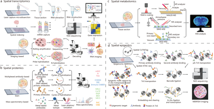
This section provides a comprehensive overview of the principles, procedures, and categorizations of spatiotemporal multi-omics, which includes spatiotemporal transcriptomics (STT), spatiotemporal proteomics (STP), spatiotemporal metabolomics (STM), and spatiotemporal epigenomics (STE). The introduction previously in this paper establishes a foundational understanding of complex organizational structures on a grander scale and across more elevated dimensions. Spatiotemporal multi-omics can be broadly classified into two principal categories. The first category involves pseudo-temporal analysis, which leverages spatial multi-omics technologies (Fig. 2). This approach entails the systematic collection of tissue samples across a series of temporal stages, followed by meticulous analysis at multiple, distinct time points. Such a method enables a nuanced comprehension of both temporal and spatial fluctuations within the tissue matrix. The second category involves the application of authentic spatiotemporal techniques, which require the labeling of specific intracellular molecules. Subsequent longitudinal tracking allows for the observation and characterization of these molecules' dynamic trajectories through time and space. These approaches collectively enhance our understanding of cellular behavior in complex biological systems. Crucially, these innovative methodologies hold immense potential in unraveling the intricate details underlying the processes of aging and regeneration, thereby driving forward our understanding and fostering the development of groundbreaking therapeutic strategies.
The multi-dimensional approach of spatial multi-omics. a Spatial transcriptomics. b Spatial proteomics. c Spatial metabolomics. d Spatial epigenomics. ISS in situ sequencing, ISH in situ hybridization, TSA tyramide signal amplification, MICS MACSima imaging cyclic staining, CODEX co-detection by IndEXing, IMC imaging mass cytometry, MIBI multiplex ionbeam imaging, DESI desorption electrospray ionization, SIMS secondary ion mass spectrometry, MALDI-MS matrix-assisted laser desorption/ionization mass spectrometry, NGS next-generation sequencing, MERFISH multiplexed error-robust fluorescence in situ hybridization, CUT&Tag cleavage under targets and tagmentation, ATAC-seq assay for transposase-accessible chromatin sequencing, mIHC multiplex immunohistochemistry, DEI diffraction enhanced imaging, MELC multi-epitope-ligand cartography, CyCIF cyclic immunofluorescence, MxIF multiplexed immunofluorescence, IBEX Iterative bleaching extends multiplexity, MSI mass spectrometry imaging. 4i iterative indirect immunofluorescence, mIHC multiplex immunohistochemistry, Immuno-SABER immuno-signal amplification by exchange reaction
Overview of STT
The widespread application of single-cell RNA sequencing (scRNA-seq) technology has yielded valuable insights into the diversity of cellular composition and gene expression status in tissues, enabling the resolution of temporal changes through sampling at distinct time points [23,24,25, 32,33,34,35,36]. It is crucial to recognize that gene expression exhibits both temporal and spatial heterogeneity. The process of scRNA-seq often involves enzymatic digestion or mechanical separation of cells into suspensions before sequencing. This procedure results in the loss of original location information and disrupts the cellular communication network, thus hindering our understanding of cellular interactions with neighboring cells and extracellular matrix. To overcome these limitations, the advent of STT technology emerged as a promising solution. STT aims to provide gene expression profiles while preserving spatial context and concurrently offering data on the temporal dimension, enabling a more comprehensive exploration of cellular interactions within the tissue microenvironment [37,38,39,40].
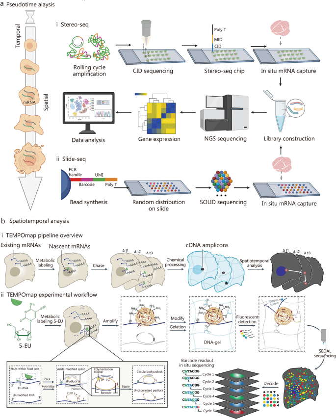
STT approaches, developed in recent years, provide a more extensive exploration of the temporal and spatial dimensions of cellular interactions within tissues. These can be broadly divided into two categories. The primary methodology employs a pseudotemporal analysis based on spatial transcriptomics techniques. It involves the systematic collection of tissue sections across various temporal stages. By conducting detailed analyses of these sections at multiple discrete time points, the method elucidates spatiotemporal variations within the tissue matrix. Presently, this pseudotemporal approach is widely applied. Conversely, the secondary methodology employs an authentic STT technique. This approach necessitates the labeling of specific intracellular substances, followed by longitudinal tracking to discern the dynamic spatiotemporal shifts they undergo. A detailed exposition of these two methodologies is presented in the subsequent sections (Fig. 3 [41,42,43]).
Integrating pseudotemporal analysis with spatiotemporal transcriptomics. a Spatial transcriptomic analyses at different time points. (i) Workflows of Sereo-seq. Reprinted with permission from [42]. Copyright © 2022. Published by Elsevier Inc. (ii) Workflows of Slide-seq. Reprinted with permission from [41]. Copyright © 2019, The American Association for the Advancement of Science. b Spatiotemporal transcriptomic analysis using 5-ethynyl uridine (5-EU) metabolic markers. (i) TEMPOmap pipeline overview: the process involves collecting and in situ sequencing nascent RNAs from various time points, followed by comprehensive spatiotemporal RNA analysis. (ii) TEMPOmap experimental workflow: the procedure starts with labeling cells using 5-EU. Three distinct probes - splint, primer, and padlock - are combined with cellular mRNAs, leading to enzymatic cDNA amplicon generation from each padlock sequence. These amplicons are integrated into a hydrogel network, secured by a specially designed acrylic component (depicted in blue). The resulting composite structure of DNA and hydrogel is visualized as blue undulating lines. Each amplicon, carrying a unique five-base barcode, undergoes sequential decoding through a six-stage process known as SEDAL fluorescence. This multiplexed RNA quantification approach precisely reveals gene expression patterns in nascent subcellular locations. Reprinted with permission from [43]. Copyright © 2023, Published by Springer Nature. CID coordinate encoding, MID molecularly encoded, NGS next-generation sequencing, PCR polymerase chain reaction, SOLID sequencing by oligonucleotide ligation and detection, SEDAL sequencing with error-reduction by dynamic annealing and ligation, cDNA complementary DNA, UMI unique molecular identifier
Pseudotemporal-based STT
We begin by presenting a brief overview of spatial transcriptomics (Fig. 2a), given that pseudotemporal analyses are fundamentally reliant on these techniques. Spatial transcriptomics technologies can be categorized into two types based on sequencing throughput. The first category includes low-throughput spatial technologies such as microdissected gene expression technologies [44,45,46,47], in situ hybridization technologies [48,49,50], and in situ sequencing methods [51,52,53]. The second category encompasses high-throughput spatial technologies that utilize spatial barcodes. Examples include Nanostring [54, 55], 10× Visium [56, 57], Slide-seq [41, 58], Slide-seq V2 [57, 59], Stereo-seq [42, 60, 61], Seq-scope [62, 63], and sci-Space [64, 65]. The foundational principles and comprehensive categorization of these techniques have been extensively discussed in our recent publications [66] and elsewhere [44, 47, 67,68,69,70,71]. The primary principles and steps are succinctly summarized (Fig. 3a [41, 42]). This section focuses on providing an overview of high-throughput spatial technologies, with a particular emphasis on Stereo-seq [42, 61, 72,73,74]. Random barcode sequences are incorporated into DNA nanoballs (DNBs) which are then positioned on a modified chip using photolithographic etching techniques. Subsequently, the array undergoes micro-graphically treatment with primers and sequencing, revealing the sequence of each etched DNB. A data matrix containing the coordinate encoding (CID) for each etched DNB is obtained. Molecularly encoded (MID) and oligonucleotide-containing polyp sequences are then bound to each position through hybridization with the CID. Frozen sections of fresh tissue are placed onto the chip’s surface for fixation, permeabilization, reverse transcription, and amplification, aimed at capturing polyA-tailed RNA from the tissue. The amplified complementary DNAs (cDNAs) serve as templates for library preparation and co-sequenced with CID. In standard data analysis, the 1 cm × 1 cm chip incorporates 400 million DNBs, a result of amplifying [61]. The Stereo-seq technology overcomes the limitations of previous spatial transcriptomic methods by simultaneously enhancing resolution, gene capture efficiency, and field of view. By introducing a novel combination of DNB patterning and in situ, RNA capture, Stereo-seq establishes a new standard for generating high-resolution, comprehensive STT atlases, holding promise for application in the realms of aging and regeneration research. It is expected to uncover potential regulatory mechanisms underlying age-related changes and tissue regeneration, thus advancing our understanding of aging and regeneration. Moreover, this technology will offer a framework for targeted interventions in age-related diseases and regenerative medicine.
Spatiotemporal-based STT
The authentic STT involves the tagging of individual cells or distinct intracellular components to observe their spatiotemporal dynamics. Presently, metabolic labeling of mRNA is instrumental in distinguishing newly synthesized mRNA molecules from those synthesized earlier. Due to advancements in biochemical methodologies, a range of RNA metabolic labeling techniques have been integrated into scRNA-seq. These methods enable the discrimination between “old” and “new” RNAs [75]. Commonly, modified exogenous nucleotides, such as 4-thiouridine (s4U) [76] and 5-ethynyl uracil [77], are employed for labeling newly transcribed RNAs. Through oxidative nucleophilic substitution, these exogenous nucleotides can be converted into different nucleotides, introducing base mutations in cDNA. This process facilitates the differentiation of “old” and “new” RNAs based on mutation sites in subsequent sequencing. Integrating the RNA metabolic labeling with high-throughput scRNA-seq, Qiu et al. [78] developed metabolic labeling-based single-cell RNA tagging sequencing (scNT-seq) using droplet microfluidic technology. In this technique, cells labeled with s4U are encapsulated in droplets along with barcoded beads. Following cell lysis, the beads capture both pre-existing and s4U-labeled newly transcribed RNA. The s4U on the beads is then chemically converted to cytosine derivatives, allowing base-pair with guanine during transcription. This process enables the location and information of new transcripts to be inferred from sequencing reads showing T to C substitutions. Although scNT-seq provides high-throughput temporal information on cellular mRNAs, it does not offer detailed transcriptional dynamics on an hourly scale.
To enhance understanding of the temporal dynamics in gene expression, Rodriques et al. [79] developed an innovative RNA timestamping technique. This method is designed to ascertain the “age” of individual RNA molecules and monitor historical shifts in gene expression within single cells. Utilizing an adenine-rich RNA template with an MS2 binding site, this RNA timestamp serves as a substrate for the enzyme adenine deaminase ADAR. In this methodology, the catalytic domain of ADAR is fused with the MS2 capsid protein. This fusion allows ADAR to specifically target and edit adenine within the MS2 binding site, leading to adenine-to-inosine (A-to-I) mutations. As ADAR binds to the RNA timestamps over time, these A-to-I edits accumulate, providing a reliable measure of the age of RNA molecules on an hourly basis. Additionally, a molecular biology protocol integrating this timestamping technique with droplet-based RNA sequencing was developed. This combination demonstrates the compatibility of the timestamping system with advanced, high-throughput single-cell transcriptomic sequencing methods. Applying this methodology enables the discernment of transcriptional changes at specific time intervals, revealing cellular diversity and applying these insights to various biological systems. This approach not only enhances understanding of transcriptional processes but also holds potential for tracking stimulus-specific responses. However, current metabolic RNA labeling methods, while enabling temporal analysis of nascent single-cell transcriptomes, lack essential spatial resolution. Although live cell imaging can track intracellular RNA trajectories, visualizing multiple transcripts concurrently remains a formidable challenge [80, 81]. There is a pressing need for highly multiplexed sequencing techniques that offer both spatial and temporal resolution, capable of effectively tracing nascent mRNA from its inception to its conclusion at subcellular and single-cell levels.
In response to this need, Ren et al. [43] developed the TEMPOmap method, designed for tracing the spatiotemporal progression of the nascent transcriptome at subcellular resolution. TEMPOmap employs the selective amplification of metabolic-labeled and pulse-labeled nascent transcriptomes, combined with advanced three-dimensional (3D) in situ RNA sequencing within hydrogel cellular scaffolds. The introduction of pulse-tracking markers enables simultaneous tracking of multiple genes throughout their RNA lifecycle, capturing vital kinetic parameters such as transcription, decay, nuclear export, and cytoplasmic translocation rates. Analysis of these spatiotemporal metrics reveals that different mRNAs undergo distinct regulation at various stages of the RNA life cycle and during cell cycle phases, influencing gene functionality. TEMPOmap represents a novel method for spatiotemporally resolved transcriptomics, utilizing metabolically labeled RNA along with a triad of probe sets: a splint DNA probe, a padlock probe, and a primer-probe. The splint DNA probe establishes a covalent bond with labeled mRNA via copper(I)-catalyzed azide-alkyne cycloaddition. Simultaneously, the padlock probe identifies the mRNA target and can loop when adjacent to the splint DNA probe on the same RNA molecule. In situ, primer probes amplify these circular padlocks through rolling circular amplification, leading to the formation of cDNA nanospheres or amplicons. This amplification occurs only for mRNAs that simultaneously bind all three probes, ensuring selective detection of labeled mRNA populations. To facilitate highly multiplexed transcriptome detection, in situ-generated cDNA amplicon libraries were embedded within hydrogel matrices. This was followed by several rounds of fluorescence imaging. Subsequently, the genes encoded by barcodes were deciphered using sequencing for error reduction by dynamic annealing and ligation (SEDAL). This approach was thoroughly tested on human cells, encompassing 991 genes, which displayed diverse spatial and temporal RNA expression patterns (Fig. 3b [43]).
As a key exemplar in the realm of spatiotemporal transcriptome research, TEMPOmap stands out for its innovative approach. This advanced technology in single-cell transcriptomics enables the simultaneous analysis of RNA with remarkable subcellular and temporal precision. It proficiently demonstrates TEMPOmap’s capability to trace the subcellular distribution and cytoplasmic translocation of transcripts over time, thereby providing a comprehensive insight into RNA subcellular dynamics at the single-cell level. Moreover, the observed strong correlation between the dynamic patterns of RNA and the molecular function of genes suggests that a functionally driven regulation of the RNA lifecycle has likely evolved to manage spatiotemporal gene expression with both precision and efficiency [82]. The validation of TEMPOmap was conducted across various cell types, including human induced pluripotent stem cell-derived cell cultures and primary cell cultures. This process illuminated the cell type-specific regulation of RNA dynamics [43]. While the kinetics of RNA generally mirror the inherent characteristics of genes grouped by molecular function, the kinetic behavior of genes crucial to specific cellular functions is notably influenced by the cell’s state and type. It is important to recognize that TEMPOmap may display sequence bias and necessitates the use of uridine analogs for metabolic labeling and the formulation of DNA probes.
An insight into STP
Traditional proteomic analyses of bulk tissues often fail to the spatial distribution and cellular specificity due to tissue homogenization, resulting in merely an average representation of protein expression levels in the mass spectrometry signal [82]. Conversely, advanced spatial proteomics provides a precise delineation of protein expression profiles across varied cells and tissue regions [83]. The integration of spatial, cell-type, and proteomic data offers critical insights into tissue spatial microenvironments. The integration facilitates the identification of accurate biomarkers and elucidates novel functional pathways [84]. While spatial transcriptomics effectively captures spatial data through methods like multiplexed fluorescence in situ hybridization or sequencing, inferring protein expression from transcriptomic data is a challenging endeavor [17]. The relationship between mRNA and its resulting protein is intricate and non-linear, often leading to discrepancies between mRNA and protein expression levels [85, 86]. Notably, transcript expression tends to exhibit more variability compared to protein expression, with proteins generally presenting lower variation coefficients relative to their corresponding mRNAs [87]. Direct spatial proteomic measurements thus provide a more authentic depiction of cellular functions and states.
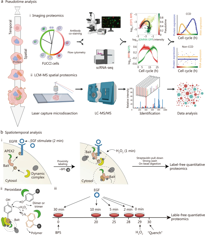
STP unveils unparalleled perspectives on protein dynamics, localization, and interactions across both spatial and temporal dimensions. It offers deeper insights into cellular processes than what can be gleaned from spatial or bulk proteomics alone [88, 89]. STP can be categorized into two primary methodologies, each characterized by its distinct approach to in vivo protein labeling for tracking intricate spatial and temporal dynamics (Fig. 4 [90, 91]). The first approach utilizes pseudotemporal analysis, where spatial proteomics techniques are employed to meticulously examine tissue sections at defined temporal intervals. Fortified by algorithms specifically designed for pseudotemporal analysis, this method extrapolates protein behaviors across the temporal and spatial spectra. Conversely, the second methodology is rooted in genuine STP. This sophisticated approach involves the in vivo tagging of targeted proteins localized within specific subcellular organelles, utilizing biotin in a live cell environment. The overarching objective here is to astutely monitor and decipher the nuanced alterations in these proteins across the intertwined dimensions of space and time [90]. While the basics of pseudotemporal analysis are well-established in spatial proteomics literature, our primary focus in this discourse is on the latter, the authentic aspect of STP. An in-depth overview of spatial proteomic methodologies is essential before delving into STP, as STP is intrinsically built upon the foundational concepts and techniques of spatial proteomics.
Temporal pseudotemporal analysis and spatiotemporal proteomic analysis. a Spatial proteomics analyses at different time points. (i) Spatial proteomic analysis uses U2OS FUCCI cells with dual fluorescent cell cycle indicators (CDT1 in G1 marked by red RFP and GMNN in S and G2 highlighted in green GFP). This system provides insights into cell cycle behavior, particularly during the G1 – S transition, where both markers are active, creating a distinct yellow hue. Using a polar model for RFP and GFP intensity, the cell cycle’s progression is streamlined into a linear format, facilitating the comparison of independent RNA and protein expression measurements aligned by individual cell pseudotime. Reprinted with permission from [91]. Copyright © 2021, Published by Springer Nature. (ii) LCM-MS spatial proteomics: utilizing laser capture microdissection followed by mass spectrometry (LCM-MS) for spatial proteomic analysis. b Spatiotemporal proteomics based on proximity labeling. (i) BP5 proximity proteomics: studies the adapter protein interactome response to epidermal growth factor (EGF) stimulation in living cells using BP5-based proximity labeling. (ii) Peroxidase-catalyzed proximity labeling: uses peroxidases for proximity labeling in both living cells and in vitro settings. (iii) Proximity proteomics workflow: involves a five-time-course EGF stimulation in HeLa cell lines stably expressing APEX2-FLAG-STS1. After EGF stimulation, cells are labeled with BP5 as indicated in the study design. Reprinted with permission from [90]). Copyright © 2021, Published by Springer Nature. CCD cell-cycle-dependent, scRNA-seq single-cell RNA sequencing, CDT1 chromatin licensing and DNA replication factor 1, RFP red fluorescent protein, GMNN geminin, GFP green fluorescent protein, EGFR epidermal growth factor receptor
Spatial proteomics
Spatial proteomics, an advanced field of study, utilizes a range of techniques, such as immunohistochemistry, immunofluorescence, mass spectrometry, and cytometry, to delineate protein distributions across scales ranging from whole tissues to subcellular entities [92, 93]. Navigating these techniques involves careful consideration of trade-offs, including spatial resolution, analytical depth, molecular and cellular throughput, as well as data acquisition durations [94, 95]. The techniques can be broadly categorized into two main groups: multiplexed antibody-based approaches and mass spectrometry-based methods, depending on the use of antibodies or other specific conjugates (Fig. 2b and Table 1 [96,97,98,99,100,101,102,103,104,105,106,107,108,109,110,111,112,113,114]).
Multiplexed antibody-based techniques employ various antibody labeling mechanisms, including fluorophores, metal markers, and DNA barcodes [94]. Each labeling method has its advantages and disadvantages, necessitating the use of validated antibodies for accuracy. Notable fluorescent techniques include SWITCH [96], multiplex immunofluorescence [97], tissue-based circular immunofluorescence [98, 99], and iterative bleach extended multiplexing [100], each distinguished by its approach to fluorescence signal removal. Indirect methods include the iterative indirect immunofluorescence [101] and the OPAL system [102]. Metal-based imaging methods, such as imaging cytometry [103, 104] and multiplex ion beam imaging by time of flight [105], utilize metal-coupled antibodies and mass spectrometry for detection. DNA barcoded techniques like DNA exchange imaging [106], CODEX [107, 108], and immuno-SABER [109], address the spectral limitations of fluorescence and bolster multiplexing capabilities. Despite the efficiency and versatility of DNA barcoding, meticulous antibody validation remains imperative.
The mass spectrometry-based method offers an alternative to antibody-based techniques. In the matrix-assisted laser desorption/ionization (MALDI) method, a pulsed laser ionizes biomolecules and peptides with near single-cell resolution (10 – 50 μm) [110]. Initially, surface proteins undergo in situ digestion to provide a peptide proteome representation. While MALDI offers high-resolution analysis of biological molecules and clarifies molecular pathways in aging and regeneration, it faces challenges such as sample degradation and matrix interferences [111]. Integrating antibody-based imaging with proteome characterization promises deeper tissue biology insights. The deep visual proteomics concept fuses AI-guided imaging with ultra-sensitive mass spectrometry-based proteomics, enhancing cell phenotype identification [112, 113]. Additionally, expansion proteomics (ProteomEx) enables high-resolution proteome profiling, recently identifying proteins in Alzheimer’s disease (AD)-afflicted mouse brains [114], marking advancements in spatial proteomics.
Pseudotemporal-based STP
The initial pseudotemporal analysis presented herein focuses on instances of single-cell proteomics methodologies applied across various phases of the cell cycle. Cell division is intricately controlled by specific proteins, encompassing their presence, and activity. These proteins are meticulously regulated in both temporal and spatial dimensions through mechanisms such as transcriptional regulation, post-translational modifications, and protein degradation [115, 116]. While traditional studies of the cell cycle have concentrated on cell populations, a deeper understanding of the intricate interplay between the cell cycle, senescence, and regeneration is of paramount importance [117]. With the progression of aging, there is an increasing dysregulation of the cell cycle, leading to reduced cellular senescence and regenerative capabilities [118]. This imbalance can precipitate age-related tissue dysfunction and the onset of diseases [119]. Comprehending and effectively manipulating the cell cycle within the contexts of senescence and regeneration are imperative for developing interventions that enhance tissue repair and delay the adverse effects of aging. Consequently, the cell cycle stands as a pivotal nexus linking the domains of aging and tissue regeneration. Previous studies have notably enhanced our comprehension of the cell cycle [115, 116], but technical constraints have limited investigations into the variability of protein expression at the single-cell level. The advent of single-cell analyses has opened novel avenues for cell cycle research. Mahdessian et al. [91] employed single-cell proteomics, coupled with single-cell transcriptomics, to identify 1180 proteins expressed in U2OS cells out of a total of 2193 proteins characterized by cellular heterogeneity. This study involved customizing specific antibodies for these proteins. Through large-scale immunostaining and systematic antibody specificity validation, single-cell proteomic data were obtained, unveiling a total of 539 cell cycle-dependent proteins, with 301 proteins previously unassociated with the cell cycle, constituting 56% of the discoveries (Fig. 4a [91]). This groundbreaking study established precise temporal expression profiles, tagged numerous proteins that play pivotal roles in proliferation, and marked the first temporal and spatial mapping of human proteome heterogeneity, systematically identifying cell cycle-associated proteins with heterogeneous expression at both mRNA and protein levels.
Spatiotemporal-based STP
The authentic STP involved the in vivo labeling of target proteins within distinct subcellular organelles using biotin in live cells, enabling the monitoring of their temporal and spatial dynamics. A recent study by Tian’s research team introduced two highly selective proximity labeling proteomics techniques [90]. These approaches revealed the spatiotemporal dynamics of interacting proteomes with remarkable temporal resolution at the subcellular level [90]. Specifically, a set of unique biotin analog probes was engineered to modulate the labeling efficiency of APEX2, thereby finely tuning the selectivity of protein complex labeling within live cells. This investigation involved the design of twelve APEX2 substrate probes, each incorporating varying electron-donating and electron-withdrawing groups strategically positioned around the phenolic structure of biotinol. These probes enable the capture of transient and weakly interacting protein complexes within living cells. A comprehensive series of in vitro and in vivo tests aimed at assessing labeling efficiency and selectivity culminated in the identification of two novel biotin analog probes, BP5 and BN2. Notably, both BP5 and BN2 probes demonstrated superior reactivity and selectivity for in vitro protein complex labeling within live cells compared to conventional biotinophenol probes (Fig. 4b [90]). This enhanced specificity in protein complex labeling is primarily attributed to their reactivity and pronounced capacity to form dimers, trimers, and even multimers, thus enabling efficient labeling of intricate protein assemblages within a confined spatial domain while reducing self-quenching. This technological advancement not only promises enhanced accuracy in deciphering protein complexes within live cells but also furnishes robust tools and methodologies for proteomic inquiries, especially in exploring intricate biological mechanisms governed by spatiotemporal dynamics at a detailed level.
STM
Genomics typically predicts potential outcomes, proteomics elucidates ongoing processes, and metabolomics reveals past events, thereby underscoring the profound capability of metabolomics to directly and precisely characterize the terminal state and phenotype of organisms [120, 121]. STM, compared to spatial metabolomics and conventional bulk metabolomics, is particularly notable in the realms of aging and regeneration research [122, 123]. STM offers precise tracking of temporal dynamics, enabling the pinpointing of transient regenerative events and distinguishing age-related metabolic changes within specific cellular compartments. This enhances our understanding of aging and regeneration with a finer level of detail. Notably, the biosynthesis, accumulation, and catabolism of metabolites in organisms exhibit a highly precise spatiotemporal distribution. The physiological functionalities of organisms are intricately intertwined with the spatial distribution of metabolites within tissues, extending even to the level of individual cells [24, 124]. Therefore, unraveling metabolite heterogeneity in both temporal and spatial dimensions is pivotal in comprehending the intricate physiological and pathological alterations occurring in organisms. Consequently, the emergence of spatial metabolomics, integrated with advanced imaging techniques, has provided a preliminary means to visualize metabolites in biological specimens, addressing our quest for metabolite visualization since the onset of the current century [125, 126]. Contemporary STM primarily relies on spatial proteomics methodologies, where tissue sections are analyzed at multiple time points, followed by the application of pseudo-spatiotemporal analysis algorithms to deduce the metabolite status across both temporal and spatial dimensions. In the following section, we will first introduce the spatial metabolomics approach, and then illustrate a representative case of STM through pseudotemporal analysis.
Spatial metabolomics
Spatial metabolomics, focusing on understanding the spatial distribution and organization of metabolites (small molecules involved in cellular processes) within biological systems such as organisms, tissues, or cells, employs mass spectrometry as a powerful method for the concurrent analysis of proteins, natural products, and metabolic derivatives [127,128,129,130]. Traditional mass spectroscopy techniques, lacking spatial information, have been supplemented by mass spectrometry-based imaging strategies using different ionization methods [131]. These methods include MALDI mass spectrometry [132, 133], desorption electrospray ionization (DESI) [134, 135], and secondary ion mass spectrometry (SIMS) [134, 135]. These techniques enable label-free detection and mapping of a wide array of metabolites, including small molecules, lipids, peptides, organic compounds, and elemental ions, and mapping within cells and tissues (Table 2 [123, 124, 132,133,134,135,136,137,138,139,140,141,142,143,144,145]). For instance, atmospheric pressure MALDI achieves up to 2 μm resolution [138] , Space-MALDI allows metabolic profiling at single-cell scales [137], and transmission-mode MALDI-2 (t-MALDI-2) reaches a resolution of 1 – 2 μm [138] for detecting phospholipids and certain biomolecules. DESI, operational under ambient conditions [139,140,141], achieves 50 – 200 μm resolution, while its variations like nanoDESI reach 10 – 15 μm [142]. SIMS-based techniques like time-of-flight (TOF)-SIMS [143] and 3D OrbiSIMS [144, 145] provide resolutions as low as 1 μm and 0.3 μm, respectively. Despite some challenges, these methods illuminate intricate molecular landscapes in tissues. Additionally, the spatial single nuclear metabolomics (SEAM) method has been introduced to tackle challenges related to segmentation and representation in SIMS data [124]. SEAM ensures the preservation of the sample’s native state through rapid and minimalistic processing, offering in situ metabolic fingerprints and individual nuclei clustering [124].
Pseudotemporal-based STM
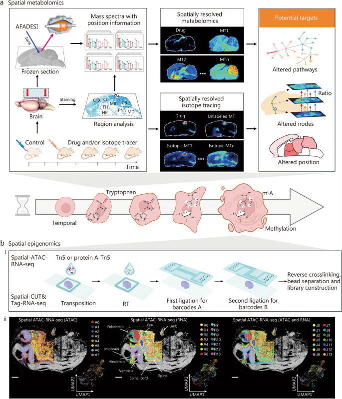
Built upon spatial proteomics methodologies, the analyses conducted at these intervals enable a refined mapping of metabolite fluctuations, enhancing our understanding of their dynamics within biological systems. We delve into an in-depth discussion of this pseudo-STM approach, drawing from recent literature. Notably, Jin et al. [123] recently employed their novel air flow-assisted desorption electrospray ionization (AFADESI)-MSI technique, merging spatially resolved metabolomics with isotope tracer analysis. Their extensive study not only examined the action mechanism of the sedative-hypnotic drug YZG-331 but also performed a multi-target analysis using an established mass spectrometry imaging method. This investigation provided a systematical exploration of both the spatial and temporal distribution of endogenous metabolites within specific microregions of the rat brain following YZG-331 administration, showcasing the sensitive and comprehensive capabilities of AFADESI-MSI (Fig. 5a [123]). The study identified functionally relevant metabolites associated with the drug action of YZG-331, localizing them within two metabolic pathways. Analysis of the “glutamine-glutamate-gamma aminobutyric acid (GABA)” metabolic pathway and isotopic glucose tracer analysis indicated a significant increase in the GABA/glutamate ratio in the hypothalamus, suggesting an enhanced glutamate decarboxylase activity post-administration of YZG-331. Furthermore, examining the “histidine-histamine-1-methylhistamine” metabolic pathway, coupled with isotopic histamine tracer studies, mainly attributed to increased peripheral histamine penetrating the pineal gland. Additionally, 1-methylhistamine levels significantly increased in the thalamic and hypothalamic regions post-administration.
Temporal pseudotemporal analysis of spatial metabolomics and epigenomics. a Spatial metabolomics analyses at different time points: this section discusses an integrated approach using mass spectrometry imaging (MSI)-based spatiotemporally resolved metabolomics combined with isotope tracing to elucidate the multifaceted targets of central nervous system drugs. The analysis spans various brain regions, including the metabolite (MT), thalamus (TH), pineal gland (PG), hypothalamus (HP), midbrain (MB), cerebellum (CB), cortex (CTX), corpus callosum (CC), pons (PN), and medulla (MD). Reprinted with permission from [123]. Copyright © 2022, Chinese Pharmaceutical Association and Institute of Materia Medica, Chinese Academy of Medical Sciences. Production and hosting by Elsevier B.V. b Spatial epigenomic analyses at different time points. (i) Schematic workflow: this part delineates a structured workflow for spatial epigenomic analysis. (ii) Spatial distribution and uniform manifold approximation and projection (UMAP) analysis: the spatial distribution and UMAP of all clusters are presented for assay for transposase-accessible chromatin (ATAC), RNA, and ATAC and RNA data. The alignment of these clusters with tissue imaging demonstrates that the spatial clusters correspond precisely with anatomical regions. The pixel size for this analysis is set at 50 µm, with scale bars representing 1 mm. Reprinted with permission from [146]. Copyright © 2023, Published by Springer Nature. m6A mRNA modification N6-methyladenosine, Tn5 pA-Tn5 transposase 5 pre-adapter-transposase 5, RT reverse transcription, CUT&Tag cleavage under targets and tagmentation
This research contributes vital insights into the metabolic mechanisms underlying YZG-331’s effects, potentially informing our understanding of age-related changes and regeneration processes. Further explorations in these areas could unveil novel therapeutic interventions for aging and regenerative medicine. Moreover, this study highlights the efficacy of advanced mass spectrometry-based imaging techniques, such as AFADESI-MSI, in elucidating complex biological phenomena. The ability to map endogenous metabolites both spatially and temporally within specific brain microregions enhances our understanding of drug actions. Additionally, integrating isotope tracer analysis amplifies our capacity to unravel intricate metabolic pathways, showcasing the potential of these cutting-edge analytical techniques in aging and regeneration research.
STE
Epigenetics, which refers to alterations in gene expression levels stemming from modifications in non-genetic sequences, is a subject of considerable focus [147]. Traditional epigenetic methods have predominantly utilized bulk sequencing, where cells from the acquired tissue are pooled before sequencing. These methods reflect an averaged state of all cells and may obscure the inherent heterogeneity among different cell types [148,149,150].
Technological advancements have facilitated single-cell-level epigenetic analysis, enabling researchers to isolate individual cells from the same tissue and assess their epigenetic status through sequencing [151,152,153,154]. However, this approach has its limitations, as tissue dissociation during single-cell suspension preparation may induce changes in intracellular gene expression and result in the loss of critical spatial information necessary for understanding cell function and corresponding biological mechanisms [153, 155]. To address these challenges, spatial epigenomics has emerged, encompassing two primary analysis methodologies: one based on next-generation sequencing, exemplified by the spatial cleavage under targets and tagmentation (CUT&Tag) technology [156, 157], which is the main focus of this paper; the other relying on high-resolution imaging techniques, such as multifluorescence in situ hybridization. Presently, spatial epigenomics primarily concentrates on transcriptome and proteome analysis [158], allowing for the simultaneous evaluation of spatial transcriptome and proteome profiles in the same tissue. The spatial CUT&Tag technology integrates microfluidic coding, a fundamental principle of the platform, with the CUT&Tag technology to investigate epigenetic modifications, enabling the precise localization of histone modifications. Understanding phenotypes relies on information from coupled genomic and transcriptomic analyses, but this does not tackle why identical DNA sequences exhibit diverse expression patterns in distinct cells. Integrating single-cell epigenomic analysis with transcriptomics can directly elucidate DNA epigenetic features, including DNA methylation, chromatin accessibility, and histone modifications associated with the originating transcriptomes. Current STE heavily relies on the spatial epigenomics approach, where tissue sections are examined at multiple time points. Pseudotemporal analysis algorithms are then applied to infer the status of gene epigenetic modifications in both temporal and spatial dimensions [159]. Therefore, we will begin by discussing the three frequently encountered forms of epigenetic modification: DNA methylation [160,161,162,163], chromatin accessibility [164, 165], and histone modifications [156, 166,167,168,169,170,171,172]. We will delineate the distinct characteristics of each modification and summarize the commonly employed methods for their investigation (Table 3 [156, 160,161,162,163,164,165,166,167,168,169,170,171,172]). Subsequently, a representative case study of STE research through pseudotemporal analysis will be illustrated.
These three epigenetic modifications intricately influence genomic function and exhibit cell type specificity. Simultaneously, there is a profound correlation between tissue structure and cellular function. As such, spatial epigenomics holds the potential to inaugurate a groundbreaking era within the field of advanced spatial genomics. In early 2022, Deng et al. [157] utilized the spatial CUT&Tag technology, an in-situ tissue encoding method, to pioneer a high spatial resolution analysis targeting specific histone modifications. This breakthrough facilitates a comprehensive examination of tissue development across spatial and genome-wide dimensions, elucidating epigenetic mechanisms underpinning both development and disease. Thereafter, Deng et al. [173] utilized spatial ATAC-seq technology to achieve in situ spatially resolved whole-genome sequencing of chromatin accessibility within tissues, marking a significant advancement. They further delved into epigenetic regulation by introducing combined spatial epigenome and spatial transcriptome sequencing approaches, embracing spatial multi-omics technologies [146]. Liu et al. [174] conducted spatial ATAC-RNA-seq combined with spatial CUT&Tag-RNA-seq analysis on mouse embryos, effectively distinguishing various organs within the embryos through the integration of epigenomic and transcriptomic data. This investigation focused on unraveling the differentiation trajectory from radial glia to postmitotic premature neurons, aiming to explore the intricate spatial and temporal correlations between chromatin accessibility and gene expression during embryonic development (Fig. 5b [146]). These findings underscore the potential of spatial ATAC-RNA-seq technology as a powerful tool for investigating gene regulatory mechanisms and unraveling spatiotemporal dynamics in the context of tissue development. Integration with single-cell data demonstrated the technique’s ability to attain cellular or near single-cell resolution.
Spatiotemporal multi-omics techniques in aging research
In this section, we delve into the applications of spatiotemporal multi-omics in the realm of aging research, underscoring their pivotal role in unraveling the complexities of aging. This exploration includes how STT reveals aging signatures and the dynamics of cellular senescence, how STP sheds light on protein biomarkers and interaction networks relevant to aging, how STM uncovers metabolic shifts and their correlations with age-related phenotypes, and how STE decodes DNA methylation patterns and epigenetic clocks that gauge biological age. Through the integration of these multidimensional omics datasets, researchers are positioned to gain deeper insights into the molecular foundations of aging, identify novel therapeutic targets for age-related diseases, and ultimately pave the way for interventions that promote healthy aging. The convergence of spatiotemporal multi-omics with aging research signals an exciting epoch of discovery. It brings us closer to understanding the intricate tapestry of the aging process and fuels the pursuit of extending the quality and duration of human life.
Application of STT in characterizing age-related tissue changes
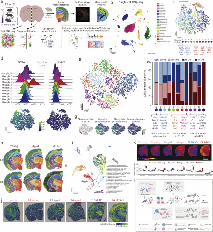
STT enables a more comprehensive investigation of cellular interactions within the aging tissue microenvironment, facilitating the discernment of dynamic changes in gene expression patterns over time and across various spatial regions within tissues. This approach provides insights into how cells communicate and adapt in the context of aging, revealing the complex molecular mechanisms driving age-related alterations in tissue structure and function [175]. STT in aging research primarily employs the pseudotemporal-based approach, which involves sequencing tissues organized at various time points using spatial transcriptomics techniques. For instance, Hahn et al. [176] employed spatial transcriptomics combined with single-cell sequencing to map the spatiotemporal transcriptome of the aging mouse brain comprehensively. Their detailed investigation revealed pronounced regional disparities in glial cell senescence, particularly within cerebral white matter glial cells, and identified specific cerebral regions responsive to regenerative interventions. Another study employed spatial multi-omics combined with single-cell sequencing across different age points to investigate the impact of apolipoprotein E (APOE) genotypes on aging, inflammatory responses, and amyloid reactions [177]. This study highlighted the role of the microglial subpopulation (Mi_6) in APOE4 carriers and senescent AD model groups, revealing how APOE4-associated microglia promote inflammation through regulatory pathways, leading to chronic neuroinflammation (Fig. 6a-g). Second, spatial transcriptomics, revealed the prevalence of PIGhigh/OLIGlow in APOE4 brains, suggesting complement activation, aberrant synaptic pruning, and disrupted axonal myelin sheath formation, which perpetuates neuroinflammation and hinders lipid metabolism (Fig. 6h-j). Furthermore, spatial metabolomics techniques identified a region in APOE4 brains associated with lipid metabolism (Fig. 6k), elucidating regulatory mechanisms making certain brain regions more susceptible to neurodegeneration in APOE4 carriers. Other studies, such as those by Stoeger et al. [178] have investigated the molecular aspects of aging by analyzing transcriptomic data from multiple studies, finding that changes in transcript length are associated with longevity. Spatial transcriptomics has been instrumental in uncovering the complex mechanisms underlying age-related changes in various tissues. Russ et al. [179] utilized this technique to examine transcriptomic changes in young and aged mouse ovaries, identifying cell-specific mechanisms that contribute to age-related fertility decline. Building on this approach, Ståhl et al. [19] mapped gene expression patterns in aged brain tissue at different times to reveal spatially distinct changes associated with aging, providing insights into the temporal dynamics of gene expression. Further demonstrating the utility of spatial transcriptomics, Asp et al. [44] characterized age-related heterogeneity within tissues, showing how different cells respond to aging processes. Besides, Kiss et al. [180] employed spatial transcriptomics to pinpoint regions in the aging mouse where senescent cells accumulate, leading to the development of inflammatory foci. This accumulation may impact age-related cognitive decline and dementia, linking cellular senescence to specific pathological outcomes in aging brains. Additionally, the utilization of spatial transcriptomic techniques and statistical methods has significantly advanced our understanding of spatial gene expression patterns, cellular senescence, and age-related processes. The introduction of Giotto by Dries et al. [181] marked a significant enhancement in analyzing and visualizing spatial transcriptomic data through a comprehensive, flexible, robust, and open-source pipeline. This development set the stage for further innovations, such as SPARK by Sun et al. [182], a statistical method specially designed to identify spatial expression patterns in spatially resolved transcriptomic, advancing our capability to interpret complex data landscapes. Building on these analytical advancements, Zhao et al. [183] introduced BayesSpace, a Bayesian method that not only enhances resolution in spatial transcriptomic data but also facilitates detailed clustering analysis, allowing for finer distinctions in tissue sample studies. Concurrently, Shang et al. [184] developed SpatialPCA, which extracts low-dimensional representations of spatial transcriptomics data while preserving the inherent biological signals and spatial correlations — essential for understanding cellular senescence and spatial gene expression patterns. Further complementing these methodological innovations, LaRocca et al. [185] discovered that noncoding repetitive element transcripts accumulate with age, serving as a reliable marker of biological age. Lastly, Kasemeier-Kulesa et al. [186] bridged single-cell and spatial transcriptomics through age- and location-matched scRNA-seq and 10× Genomics Visium analyses, providing a comprehensive view of gene expression and cellular behavior in aging tissues.
Implementing spatiotemporal multi-omics in aging research. a Brain analysis in APOE3 and APOE4 mice were systematically analyzed at different life stages (3, 12, and 24 months) and under specific conditions such as inflammatory challenge [lipopolysaccharide (LPS)] and Alzheimer’s disease (AD) pathology (characterized by amyloid overexpression). b Uniform manifold approximation and projection (UMAP) classification of gene expression clusters: a UMAP analysis identified 24 distinct clusters based on canonical gene expression markers. c Microglia sub-cluster analysis using t-distributed stochastic neighbor embedding (t-SNE): t-SNE revealed the microglial sub-clusters. Key biomarkers for “homeostatic” clusters (0 and 1) and disease-associated microglia (DAM)-like cluster 6 are annotated below the respective cluster labels. d Analysis of regulon activity scores: ridge plots (left) and t-SNE plots (right) illustrate the activity scores of HIF1a and Srebf2 regulons. e Microglial sub-cluster distribution in LPS- or saline-treated E3 and E4 mice: a t-SNE plot displays the distribution of 12 microglia sub-clusters in mice treated with LPS or saline, differentiated by colors. f The allocation of experimental groups across various microglia sub-clusters is depicted through a stacked bar chart. This visualization effectively illustrates the distribution of experimental groups among the different sub-clusters of microglia. g Within E4 LPS brains, t-SNE plots have uncovered a significant upregulation in crucial carbon pathways related to energy production. This increased activity is notably prevalent in specific subclusters found in brains with the E4 genotype that have undergone LPS treatment. h Spatial transcriptomic analysis is applied to brain sections from young, aged, and amyloid-overexpressing E3 and E4 mice, offering a detailed examination of genetic expression across different brain regions and conditions. i A thorough UMAP analysis is conducted for comprehensive data interpretation. j In spatial transcriptomic plots, DAM/neurodegenerative microglia (MgND) scores are displayed for each spot, calculated via AUCell. This analysis incorporates a brain from each experimental group for comparison. k The study also includes an analysis of a specific lipid, phosphatidylcholine (16:0/18:2). Top scans illustrate the lipid’s spatial distribution in coronal brain sections, while the bottom part quantifies the average pixel intensity of this phosphatidylcholine variant across different brain regions, offering a visual and quantitative insight into its distribution. Reprinted with permission from [177]. Copyright © 2023 The Author(s). Published by Elsevier Inc. l Schematic overview of spatial genome architectures in growing (center) and senescent (gray boxes) stem cells. Reprinted with permission from [187]. Copyright © 2022, Oxford University Press. APOE apolipoprotein E, LAD-NL lamina-associated domain-nuclear lamina, iLAD inter-LAD-NL
Collectively, these studies provide valuable insights into the spatial distribution of gene expression, the impact of cellular senescence on cognitive decline and neuroinflammation, and the potential mechanisms underlying regenerative and non-regenerative healing. The development of robust pipelines and statistical methods has enabled more effective analysis and visualization of spatial transcriptomic data, facilitating a deeper understanding of cellular processes and interactions within tissues. These findings have significant implications for future research in understanding the molecular changes underlying aging and developing novel therapeutic strategies for age-related disorders.
STP and STM approaches to elucidate molecular dynamics during aging
STP has revolutionized our understanding of aging by providing a dynamic perspective, enabling the identification of key specific proteins and pathways central to age-related changes [188]. This knowledge is invaluable for developing interventions targeting the molecular drivers of aging. A recent study employing STP systematically analyzed comprehensive proteomic profiles across various brain regions in non-human primates from fetuses to neonates [189]. This approach offers valuable insights into normal brain development and informs our understanding of mechanisms underlying dysfunctions and disorders in humans, with profound implications for neuroscience research. Hosp et al. [190] utilized STP to investigate proteomic changes during the progression of Huntington’s disease (HD) in the R6/2 mouse model. This study highlighted substantial reconfiguration of the soluble brain proteome, closely associated with the emergence of insoluble aggregates as the disease advanced. The accumulation of proteins in these aggregates was linked to their expression levels and specific sequence characteristics, underscoring the role of cellular protein dysfunction in HD toxicity. STP has identified proteins associated with aging, such as telomerase [191, 192] and sirtuins [193, 194]. The aberrant modulation of protein expression and functionality during aging is recognized as a key influence on the aging phenomenon [195]. Telomerase, typically quiescent in most adult cells but active in cancer cells and stem cells, presents a promising target for potentially decelerating or even reversing the aging process [191]. Huang et al. [196] utilized spatial proteomic analyses in 4 distinct brain regions, revealing differential proteins associated with resilient populations in AD. These studies underscore the significance of STP in elucidating key protein targets in aging and forming a basis for therapeutic interventions in age-related diseases.
Similarly, STM provides a comprehensive view of metabolic changes throughout the aging process. Walker et al. [197] at the University of California produced the first spatial and temporal metabolite map of the mouse brain across the aging spectrum, from adolescence to old age. This extensive dataset, encompassing 1547 molecules across 10 brain regions and 4 developmental stages [197], highlights significant variations in metabolites with no sex correlation. Notable findings include changes in sphingolipid patterns, indicative of myelin remodeling, and metabolic pathways during aging. The study revealed weakening metabolic correlations in the cerebrum from adolescence to adulthood and reduced cerebral segregation in old age [198]. These metabolic shifts, when correlated with gene and protein brain atlases, offer insights into brain metabolism’s spatial and temporal dynamics during aging. Additionally, spatial metabolomics techniques have been employed to investigate age-related metabolic changes, revealing spatially distinct metabolic signatures of aging [199]. Integrating spatial proteomic and metabolomic data has facilitated the identification of molecular interactions and pathways involved in aging [200,201,202]. These studies highlight the importance of proteomics and metabolomics in comprehensively elucidating the molecular mechanisms of aging in both spatial and spatial dimensions, leveraging advancements in STP and metabolomic techniques. Understanding molecular dynamics within tissues aids in deciphering age-related changes, identifying potential biomarkers, and developing targeted interventions for healthy aging.
STE profiling in a spatial context to understand age-related modifications
STE has arisen as a transformative methodology, enabling the elucidation of intricate interactions among epigenetic modifications, spatial arrangement, and temporal dynamics within the context of aging. This section delves into the pivotal function of STE in enhancing our comprehension of the molecular mechanisms underlying the aging process. Drawing upon perspectives from prominent researchers and recent empirical discoveries, this section insights into the diverse applications of STE in the field of aging research. STE provides a dynamic view of how our epigenome evolves with age. By understanding these changes within a spatial and temporal framework, we gain unprecedented insights into the epigenetic basis of aging, offering potential avenues for interventions to promote healthy aging.
The aging process is characterized by a multitude of epigenetic alterations, encompassing DNA methylation, chromatin accessibility, and histone modifications [2, 3, 203]. These tightly regulated and frequently reversible modifications exert influence over gene expression and various cellular processes, ultimately contributing to the onset and progression of numerous age-associated human disorders. A plethora of enzyme systems, including DNA methyltransferases, histone acetylases, deacetylases, methylases, and demethylases, in conjunction with chromatin remodeling factors, participate in establishing and maintaining epigenetic patterns [204].
The landscape of DNA methylation in humans has undergone cumulative alterations over time [204]. Initial investigations highlighted generalized age-related hypomethylation, yet subsequent analyses unveiled specific loci, including those associated with certain tumor suppressor genes and polycomb target genes, exhibiting heightened methylation levels during aging [203]. Additionally, cells derived from individuals and mice manifesting progeria-like syndromes demonstrate DNA methylation shifts that partially mirror those observed in typical aging processes [203, 205]. However, the functional implications of these age-associated DNA methylation modifications remain elusive, primarily due to a lack of understanding regarding their temporal and spatial dynamics. STE, equipped to elucidate the distribution of epigenetic marks within cells and tissues across space and time, introduces a fresh vantage into the molecular dynamics underpinning aging. Rodriguez-Muela et al. [206] pioneered the use of spatial epigenomics to investigate how DNA methylation patterns shift with age, identifying specific epigenetic alterations that vary distinctly across different tissue regions. Building on this foundation, Zocher et al. [207] extended these analyses to brain tissue, where they mapped the spatial variability of DNA methylation changes, further detailing the epigenetic landscape of aging neural tissue. Smith et al. [208] underscored the importance of comprehending these spatiotemporal dynamics, emphasizing that a comprehensive grasp of epigenetic modifications throughout the aging process is crucial for developing targeted anti-aging therapies. Complementing these findings, Wu et al. [209] broadened the scope of the STE study by examining how the common mRNA modification N6-methyladenosine (m6A) affects primate tissue health and aging. They discovered tissue-specific m6A changes in the liver, heart, and skeletal muscle of both young and aged nonhuman primates, particularly emphasizing the susceptibility of skeletal muscle to m6A reduction during aging and highlighting the crucial role of the m6A methyltransferase-like 3 (METTL3) in maintaining muscle health. These findings shed light on the mechanisms underlying tissue aging and reveal a METTL3-m6A- nephronectin (NPNT) axis that helps mitigate muscle degeneration associated with aging.
Aging is strongly associated with both global histone loss and tissue-specific alterations in post-translational modifications of histones [3, 210]. Enhanced histone expression has been linked to extended lifespan in Drosophila, while investigations in fibroblasts from older individuals and patients with progeria have revealed increased histone H4K16 acetylation or H3K4 trimethylation, alongside reduced levels of H3K9 or H3K27 trimethylation [211]. These modifications of histones have the potential to induce shifts in transcriptional activity, disrupt cellular homeostasis, and contribute to age-related metabolic decline. Notably, the diminishment of telomeric heterochromatic markers has been demonstrated to result in telomere elongation.
In addition to DNA and histone-modifying factors, various chromosomal proteins and chromatin remodeling factors, including heterochromatin protein 1a and polycomb histones, play a role in genome-stabilizing DNA repair and senescence regulation [212]. The lowest hierarchical level of chromosomal organization involves the folding of chromatin fibers, intimately associated with chromatin recycling [187]. This recycling process, characterized by myriad DNA and histone modifications, plays a pivotal role in transcriptional regulation, determining DNA accessibility to the transcriptional machinery (Fig. 6l [187]). Modifications in these epigenetic elements result in significant shifts in chromatin configuration, encompassing widespread heterochromatin depletion and repositioning, prevalent occurrences in senescent cells. For instance, Zhang et al. [213] employed advanced epigenetic high-throughput methods, including chromatin accessibility sequencing, chromatin immunoprecipitation sequencing, limited enzyme digestion, and isotope-labeled quantitative proteomics, to unveil fundamental patterns of histone epigenetic modifications in senescent cells, elucidating the dynamics of chromatin spatial accessibility, and highlighting the significant contributions of epigenetic factors like lysine demethylase 4. The study underscores the potential targeting value of these molecules for manipulating senescence.
STE offers an innovative perspective for exploring the dynamic epigenetic alterations accompanying aging. By unveiling the spatiotemporal distribution of epigenetic marks, this paradigm enhances our understanding of the molecular mechanisms driving the aging process while delineating potential avenues for interventions aimed at fostering healthy aging.
Spatiotemporal multi-omics techniques in regeneration studies
Regeneration stands as a remarkable biological phenomenon observed across the animal kingdom, encompassing a broad spectrum of organisms, from invertebrates like planarians to vertebrates like salamanders. However, within mammals, including humans, regenerative potential remains comparatively limited, predominantly confined to select tissues, such as the liver and skin. In the realm of regenerative medicine, the integration of spatiotemporal multi-omics approaches holds great promise. Drawing upon insights gleaned from existing literature, the hallmarks of regeneration can be conveniently categorized into three principal domains: the classification of regeneration types, the underlying mechanisms governing regeneration, and the regulatory processes that orchestrate these events. Regeneration is typically classified into three types, complete, incomplete, or compensatory, depending on the extent of restoration achieved [214,215,216,217]. Complete regeneration entails the comprehensive reinstatement of a lost or damaged component, achieving both structural and functional recovery. This phenomenon is observed in organisms as diverse as planarians. In contrast, incomplete regeneration results in only partial restoration, potentially recovering some, but not all, aspects of structure or function, as seen in peripheral nerves. Compensatory regeneration, on the other hand, allows an organ or tissue to regrow to functional sufficiency without replicating the original form or achieving complete functionality. A prime example is live regeneration in various vertebrates, including humans, where hepatocytes proliferate to regain function, though not the original liver structure (Fig. 1b).
The processes governing regeneration predominantly encompass cell proliferation, migration, differentiation, and the precise patterning of regenerating tissues and organs [218, 219]. Cell proliferation serves as the foundational process, where new tissues or organs arise through cellular division and propagation [220,221,222,223]. Cell migration involves the movement of cells from one location to another, establishing the correct structural arrangement [221, 224, 225]. Cell differentiation marks the transformation of cells from an undifferentiated state into specialized cell types, such as muscle or nerve cells [220, 221]. Pattern formation of regenerating organs refers to the capacity of newly generated tissues or organs to develop in a specific sequence and arrangement during the regeneration process [226, 227]. Governing the regeneration process is an array of influential factors, encompassing genes (i.e, MSX, p53, HOX) [228,229,230,231,232,233], environmental elements (i.e., temperature) [234, 235], hormones (i.e., androgens and estrogens) [128, 129, 236,237,238,239], and growth factors (fibroblast growth factor and epidermal growth factor) [240,241,242,243]. Of particular significance, genes play a pivotal role in the regulation of cell proliferation, differentiation, and migration, intricately controlling the complex process of regeneration.
The integration of spatiotemporal multi-omics techniques into regenerative medicine holds the potential to offer invaluable insights into the intricate and dynamic molecular events governing tissue regeneration. This integration may pave the way for novel therapeutic approaches and enhance our understanding of the regenerative potential within mammals, including humans. In this review, we explore recent applications of spatiotemporal multi-omics approaches within the field of regenerative medicine, shedding light on their potential to revolutionize our understanding of regenerative processes and their therapeutic implications.
Utilizing STT in investigating tissue regeneration processes
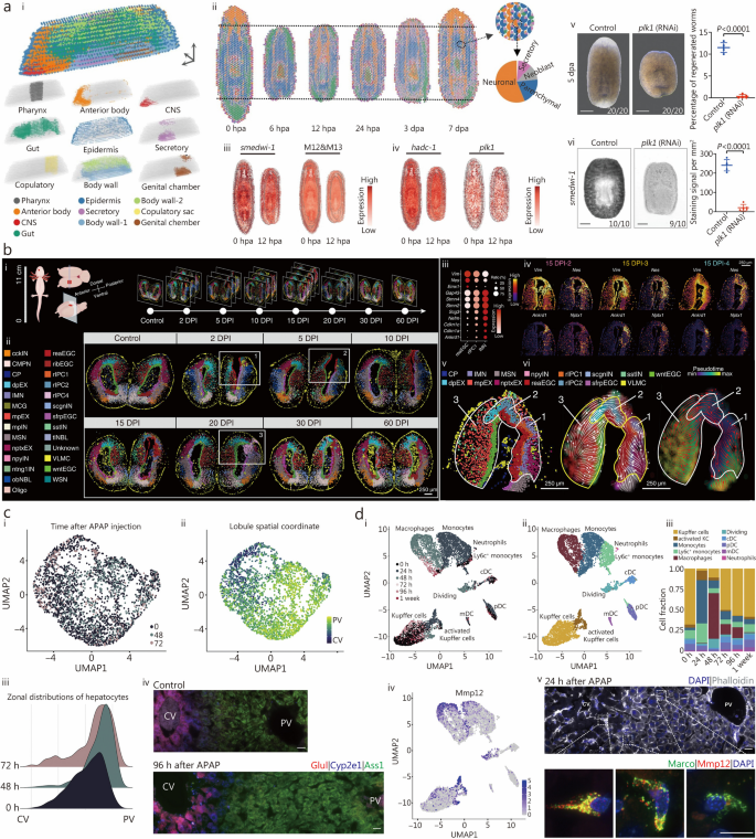
STT, integrating high-throughput gene expression profiling with spatial and temporal data, enables detailed characterization of gene expression patterns in regenerating tissues. This technique captures the spatial organization of gene expression over time, allowing researchers to identify specific cell types, signaling pathways, and molecular signatures crucial to tissue regeneration [19, 244]. It provides a comprehensive perspective on transcriptional changes during different stages of tissue repair, illuminating the dynamic cellular responses and regulatory networks involved in regeneration. For instance, Cui et al. [245] conducted a pioneering study on planarian regeneration. By merging spatial transcriptomics with single-cell sequencing, they created a 3D transcriptome atlas detailing characteristic cell distributions and gene expression patterns at six-time points, later expanded with two additional time points (Fig. 7ai). This study revealed a totipotent stem cell cluster and identified multiple gene expression patterns linked to planarian regeneration (Fig. 7aii). Using spatial module screening tools, the study pinpointed polar gene pairs crucial to wound-specific expression and to regions pre-/post-planarian. A key regulatory gene, plk1, was identified as critical in planarian formation and regeneration (Fig. 7aiii-iv), with functional analyses indicating its role as an early response element in blastocyst formation and subsequent regeneration events (Fig. 7av-vi). These insights notably enhance our comprehension of the intricate regulatory mechanism’s planarian regeneration, relevant to both specific planarian studies and broader mammalian regeneration research.
Leveraging spatiotemporal multi-omics in regenerative studies. a Planarian regeneration analysis: (i) spatial transcriptomics spots were grouped using the STAGATE method, labeling clusters based on their spatial expression patterns. (ii) The study visually tracks primary cell types’ spatial distribution during regeneration stages using pie charts in corresponding spatial transcriptomics spots. (iii) Spatial distribution mapping of smedwi-1 and neoblast-related genes provides insights into their localization. (iv) Spatial transcriptomics data at 0 and 12 hpa examines spatial expression patterns of regulatory genes hdac-1 and plk1 during early regeneration. (v) A plk1 knockdown effect in planarians at 5 dpa is illustrated in a bright-field image. (vi) WISH staining of smedwi-1 in control and plk1 knockdown planarians at 3 dpa. Reprinted with permission from [245]. Copyright © 2023 The Author(s). Published by Springer Nature. b Axolotl telencephalon regeneration research: (i) schematic illustrates sample collection and examined sections during homeostatic and regenerative phases. (ii) Stereo-seq maps cell types in axolotl telencephalon sections using various stages of regeneration. (iii) Bubble chart displays the fluctuating expression of marker genes defining cell types crucial for telencephalon regeneration. (iv) Heatmap shows spatial expression trends of essential markers within the injured area at 15 DPI-2, -3, and -4. (v) The spatial arrangement of cell types around the regeneration site is shown for the 15 DPI-4 section. (vi) RNA velocity plots illustrate predicted cellular lineage transitions in regenerating axolotl telencephalon area. Reprinted with permission from [60]. Copyright © 2022, The American Association for the Advancement of Science. c Hepatocyte study post-APAP injection: (i) UMAP visual representation of hepatocytes color-coded by time post-APAP injection. (ii) UMAP of hepatocytes color-coded by inferred spatial coordinates within the lobule. (iii) Distribution of spatial coordinates among hepatocytes for different time intervals post-injection. (iv) smFISH images of a liver lobule exhibit zonated genes at different time points post-APAP. Reprinted with permission from [246]. Copyright © 2022 Elsevier Inc. d Myeloid cell analysis post-APAP administration: (i) UMAP visualization of myeloid cell populations color-coded by time post-APAP administration. (ii) UMAP color-coded by myeloid cell types. (iii) Proportions of various myeloid cell subtypes at each examined time point. (iv) UMAP visualization of myeloid cells, colored by expression levels of Mmp12. (v) smFISH scan (top) and zoomed-in insets (bottom) of a liver lobule 24 h after APAP injection. Reprinted with permission from [246]. Copyright © 2022 Elsevier Inc. CNS central nervous system, hpa hours post-amputation, dpa days post-amputation, WISH whole-mount in situ hybridization, reaEGC reactive ependymoma cell, IMN immature neuron, rIPC1 regeneration intermediate progenitor cells 1, UMAP uniform manifold approximation and projection, APAP acetaminophen, CV central vein, PV portal vein, smFISH single-molecule fluorescence in situ hybridization, KC kupffer cells, pDC plasmacytoid dendritic cells, cDC conventional dendritic cells, mDC myeloid dendritic cells, Mmp12 matrix metallopeptidase 12, UMI unique molecular identifier
In the regeneration field, a multi-institute research team led by the Beijing Genomics Institute recently applied Stereo-seq to examine self-healing processes of brain injuries in axolotls [60]. They conducted Stereo-seq analysis on a cerebral cortex section from the salamander’s telencephalon during a 60-day post-recovery period (Fig. 7bi). This approach identified 28 distinct cell types (Fig. 7bii), revealing that morphological wound healing occurred around 30-d post-injury, with complete cell type restoration by 60-d post-injury. This study identified an additional cell type that exhibits patterns similar to those of reactive ependymoma cells (reaEGCs) and immature neurons (IMNs), and expressing markers from both categories (Fig. 7biii-iv). This cell type, termed “regeneration intermediate progenitor cells 1” (rIPC1) suggests a sequential transition of cell types (reaEGCs-rIPC1-IMNs) during regeneration. RNA velocity analysis confirmed the interconnected sequence of these cell types (Fig. 7bv-vi), providing compelling evidence for their interdependence in the regeneration process. The establishment of spatiotemporal cellular maps for salamander brain development and regeneration is crucial in understanding brain regeneration, the intricate structure of amphibian brains, and evolutionary brain structure trajectories. It also opens new avenues for clinical interventions in human tissue and organ self-repair and regeneration, while offering a valuable data repository for species evolution studies. Ben-Moshe et al. [246] conducted a comprehensive investigation into liver regeneration dynamics using spatial transcriptomics, single-cell sequencing, and bulk transcriptome sequencing. Their work elucidated the mechanisms underlying zonal hepatocyte regeneration and the supportive role of various region-specific non-parenchymal cells (Fig. 7c). They noted the downregulation of major histocompatibility complex class I and class II molecules, inhibiting adaptive immune activation during liver regeneration (Fig. 7d). This study severs as a comprehensive resource on the coordinated mechanisms of zonal liver regeneration, enriching our understanding of this complex process.
Collectively, these studies highlight the potential role of STT in regenerative medicine. Employing axolotls, planarians, and liver models, they provide invaluable insights into the mechanisms of tissue repair. By mapping gene expression changes within a spatial and temporal context, researchers can identify key genes, cell types, and pathways essential for successful regeneration. This methodology not only deepens our comprehension of regenerative phenomena but also offers valuable tools for advancing regenerative medicine research. STT is poised to unravel the complexities of tissue restoration and drive progress in regenerative therapies.
STP and STM in identifying key factors and pathways in regeneration
Recent advancements in STP and STM have yielded powerful tools for dissecting the intricate processes involved in regeneration. This section delves into the invaluable contributions of these cutting-edge technologies in identifying key factors and pathways crucial for the successful regeneration of tissue and organs. By elucidating protein expression patterns and metabolic changes, researchers can unravel the spatially restricted molecular dynamics that underlie the process of tissue repair [34, 247, 248]. This innovative approach facilitates the discovery of novel biomarkers and therapeutic targets, thereby enhancing the prospects of regeneration.
STP is geared towards elucidating the spatial distribution and temporal dynamics of proteins within regenerating tissues. This approach has played a pivotal role in unraveling the molecular mechanisms that drive regeneration across diverse model organisms. In a notable study, Mallah et al. [249] conducted a spatiotemporal microproteomic analysis to explore traumatic brain injury (TBI) over a 10-d post-injury period. Their findings showcased a restoration of the brain’s protein profile to its pre-injury state within this timeframe. Intriguingly, they also identified a pronounced upregulation of proteins associated with Parkinson’s disease (PD) in the brain’s substantia nigra as early as 3 d post-injury, hinting at a potential association between TBI and PD. This investigation, employing spatiotemporal microproteomics, provides invaluable insights for regenerative medicine. By identifying specific protein markers and tracing their trajectories immediately following injury, the study sheds light on the intrinsic regenerative capacities of the brain. The suggested connection between TBI and PD underscores the need to comprehend the broader implications of traumatic injuries, a perspective crucial for shaping regenerative and neurotherapeutic strategies. Furthermore, the employment of in vitro models to validate these findings amplifies the translational significance of the study. Huang et al. [196] examined 4 distinct brain regions such as the caudate nucleus, hippocampus, inferior temporal parietal lobule, and the middle gyrus of the superior temporal gyrus in their study involving 11 healthy controls, 12 individuals demonstrating resilience to Alzheimer’s disease (RAD), and 20 patients with AD. Their results identified 33 proteins differentially expressed in the resilient group. Notably, 5 of these proteins — platelet-activating factor acetylhydrolase Isoform 1B subunit 3 (PA1B3), testican-3 (TICN3), intercellular adhesion molecule 1 (ICAM1), immunity-related GTPase Q (IRGQ), and aldehyde dehydrogenase 1 family member L1 (AL1L1) — were uniquely associated with RAD, suggesting their potential significance in the pathogenesis of AD. This study, by highlighting reduced soluble Aβ levels and pinpointing specific proteins linked to RAD, provides crucial insights that may prove instrumental for regenerative medicine research and future therapeutic interventions for AD.
Spatiotemporal protein expression profiling also offers fresh insights into peripheral nerve regeneration mechanisms and potential therapeutic targets. Bryan et al. [250] employed a reverse phase protein microarray approach to investigate the dynamic protein expression patterns during the 28-d course of peripheral nerve regeneration in a rat sciatic nerve transection injury model. The research delves into the spatiotemporal profiles of various proteins involved in nerve regeneration, including growth factors, extracellular matrix proteins, and those related to adhesion and migration. The findings reveal distinct changes in protein expression across different segments of the regenerating nerve and suggest potential implications for future research in understanding the molecular mechanisms underpinning nerve regeneration and identifying therapeutic targets.
STM, on the other hand, centers on the study of the changes in metabolite levels and fluxes within tissues during regeneration, offering insights into the metabolic processes that underpin tissue repair. A study on liver regeneration after acute damage integrated spatial metabolomics with spatially resolved scRNA-seq to elucidate the dynamics of regeneration [246]. This integrated approach unveiled the coordinated roles of different cell types, including hepatocytes, endothelial cells, hepatic stellate cells, and macrophages, in liver tissue repair. STP and STM have emerged as indispensable tools for dissecting the complexities of tissue and organ regeneration. They provide a comprehensive view of the molecular events that transpire during the repair process. Ongoing research in this domain holds great promise for regenerative medicine, as it offers insights into potential targets for therapeutic interventions aimed at enhancing tissue regeneration.
Epigenetic regulation in spatial and temporal dimensions during tissue repair
Epigenetic modifications play a critical role in regulating gene expression and cellular identity during tissue regeneration [36, 251]. Spatial epigenetic profiling allows for the investigation of epigenetic changes within the spatial and temporal context of tissue repair [206]. By examining DNA methylation patterns and chromatin states, researchers can gain insights into the spatially defined epigenetic regulation of regeneration-associated genes and the establishment of cell fate during tissue repair. This knowledge enhances our understanding of the epigenetic mechanisms governing regeneration.
Challenges and future prospects
In the fields of aging and regenerative medicine, a host of challenges beckon, including the complexity of data integration, the imperative for high-resolution techniques, the quest to decipher temporal dynamics, the enigma of interpreting multimodal data, and the quest for standardization. The horizon, however, gleams with promise in the realms of single-cell profiling, multi-omics integration, machine learning and AI, functional validation, and precision medicine. These avenues offer profound insights, interventions, and early detection mechanisms in the relentless pursuit of unraveling aging and tissue regeneration (Fig. 8).
Challenges and future directions in spatiotemporal multi-omics research. In the domains of aging and regenerative medicine, challenges encompass complexities in data integration, the imperative for high-resolution techniques, comprehension of temporal dynamics, interpretation of multimodal data, and the establishment of standardization. Looking ahead, there is great promise in single-cell profiling, multi-omics integration, machine learning and AI, functional validation, and precision medicine. These advancements hold the potential to provide profound insights, interventions, and early detection capabilities in the study of aging and tissue regeneration. m6A mRNA modification N6-methyladenosine, AI artificial intelligence
Challenges
Data integration complexity
In the realm of aging and regeneration research, deciphering the intricate relationship of spatial and temporal factors stands as a linchpin for uncovering the core molecular mechanisms. Traditional multi-omics approaches, while offering significant molecular insights, frequently fall short in terms of spatial and temporal resolution [82, 252,253,254,255,256,257]. This limitation threatens to obscure a comprehensive comprehension of processes like aging and tissue regeneration. Consequently, spatiotemporal multi-omics techniques that integrate both spatial and temporal dimensions have gained prominence. Such integration captures the dynamic intra-tissue changes over time and discerns molecular signatures specific to various aging or regeneration phases, enriching our understanding of spatial and temporal nuances [37, 258].
However, the analysis of extensive spatiotemporal omics datasets presents distinct challenges, necessitating bespoke computational methods. These challenges include handling the data’s high dimensionality and intricate spatial structures [259, 260], as well as discerning spatial patterns and cellular diversities [261]. In response, tools such as spatial clustering algorithms and spatially constrained dimensionality reduction have been developed. For instance, the utilization of spatially constrained non-negative matrix factorization aids in identifying unique cell types and their gene expression profiles [34, 262, 263]. Moreover, the integration of diverse spatiotemporal datasets, spanning transcriptomics to epigenomics, presents a formidable undertaking [264,265,266]. These datasets often derive from different platforms, leading to disparities in data types and scales. Overcoming these demands, the creation of robust analytical tools for data harmonization and the introduction of advanced techniques, including multi-omics integration algorithms, becomes imperative. These tools are instrumental in harmonizing and deciphering the varied and intricate datasets, thereby enabling researchers to distill invaluable insights from the integration of diverse omics layers. This integrative approach stands as a cornerstone for unraveling the multifaceted aspects of aging and regeneration, providing a more comprehensive understanding of their spatiotemporal intricacies. The achievement of proficient data integration thus becomes pivotal in our pursuit of comprehending the intricate spatiotemporal intricacies of aging and regeneration.
High-resolution techniques
Obtaining high-resolution data capable of capturing dynamic changes at the cellular and subcellular levels emerges as a categorical imperative. Existing techniques, often lacking the necessary spatial and temporal granularity to dissect intricate processes during aging and regeneration, necessitate a quantum leap. The development of cutting-edge imaging and omics technologies stands as the panacea to surmount this challenge. For instance, single-cell multi-omics approaches have the potential to unveil granular insights into individual cell behaviors over time [165, 267,268,269]. Advanced imaging techniques, including super-resolution microscopy and live cell imaging, offer high-resolution spatial and temporal data [270,271,272,273,274]. Strategic investment in research and innovation to refine and expand these techniques will be critical for gaining a deeper into the spatiotemporal dynamics within these domains.
Temporal dynamics
Comprehending the temporal dynamics of biological processes takes center stage in aging and regeneration research. Current methods often provide snapshots of static states, thereby rendering the capture of dynamic changes a formidable task. In Part II, spatiotemporal multi-omics bifurcate into two main classes. The first entails pseudotemporal analysis through the utilization of spatial multi-omics, examining tissue sections from diverse time intervals. The second employs genuine spatiotemporal genomics techniques, labeling specific cellular components to investigate temporal changes. However, the pseudotemporal approach remains dominant in aging research due to its established methodology and ease of use [43, 275]. Addressing this challenge requires the conception of longitudinal and time-series experimental designs. Researchers must employ technologies that facilitate real-time monitoring and sampling of biological systems over extended durations. Additionally, computational tools for modeling and analyzing temporal data necessitate enhancement to extract meaningful patterns and identify critical transition points during aging and regeneration.
Interpreting multimodal data
The integration of multiple omics modalities, encompassing genomics, proteomics, and metabolomics, beckons the need for comprehensive frameworks for interpreting data effectively. The intricate interactions across these layers in aging and regeneration require innovative approaches to unravel intricate networks and pathways [276]. Techniques for multimodal data integration, including network-based analyses and pathway enrichment methods, must be refined to account for dependencies and interactions between distinct omics components [277,278,279,280]. Furthermore, interdisciplinary collaboration spanning biology, bioinformatics, and systems biology, is indispensable for advancing our ability to interpret multimodal data proficiently. Contemporary biotechnology enables the simultaneous measurement of multiple high-dimensional modalities (e.g., RNA, DNA accessibility, and proteins) within a single tissue sample. To comprehensively elucidate the role of gene regulation in steering biodiversity and function, a combination of diverse analytical approaches becomes imperative. However, prevailing methods primarily cater to single-task analysis, offering only a partial insight into multimodal data. Recent breakthroughs in single-cell biotechnology empower the concurrent assessment of gene expression and other high-dimensional modalities within the same cell, yielding multimodal histological data [281,282,283]. This data vantage furnishes a comprehensive view of cellular transcriptional and functional processes. Nonetheless, traditional methodologies fall short of handling such multimodal data, necessitating the development of novel approaches to fully harness their potential [277, 278]. While researchers have put forth various methods for multimodal analysis, tackling tasks such as joint group identification, cross-modal prediction, and cross-modal correlation discovery, these methodologies are typically tailored with a singular objective, posing challenges for their integration into a unified framework [284,285,286,287]. To bridge this divide, Tang et al. [288] introduced interpretable multi-task deep neural networks, demonstrating efficacy in navigating the intricacies of multimodal data analysis. The network utilizes an encoder-decoder-discriminator architecture, which is iteratively trained to perform tasks such as joint cluster identification and cross-modal prediction. Such a framework holds significant potential to accelerate the uncovering of cell-type-specific regulatory dynamics across transcriptomics and other modalities.
Standardization
The establishment of standardized protocols and data formats for spatiotemporal multi-omics data assumes paramount importance, ensuring data reproducibility and comparability across studies [289, 290]. Presently, variations in experimental protocols, data preprocessing steps, and data reporting impede data harmonization. Collaborative endeavors among researchers are imperative to define and embrace common data standards and best practices. This includes the development of metadata standards encompassing experimental conditions, sample information, and data preprocessing steps. Moreover, the establishment of data repositories and platforms adhering to these standards becomes imperative to facilitate data sharing and reproducibility within the scientific community. Standardization efforts will enhance the reliability and utility of spatiotemporal multi-omics data in aging and regeneration research.
Future prospects
Integration of additional layers such as 3D configuration and single-cell resolution
The horizon of spatiotemporal multi-omics research in the context of aging and regeneration radiates with great promise, especially within the realm of single-cell analysis. These advancements will furnish a granular perspective of individual cells as they evolve over time [291,292,293]. This approach will uncover previously veiled cellular heterogeneity and dynamics, enabling the discernment of rare cell populations, transitional states, and responses of cells to environmental cues. Single-cell spatiotemporal profiling is poised to be instrumental in unraveling the intricacies of tissue regeneration, elucidating the roles of distinct cell types, and meticulously tracking their behavior throughout the intricate process.
Moreover, the incorporation of additional layers of information, such as 3D configuration and single-cell resolution, constitutes a pivotal direction for advancing spatiotemporal omics within aging and regeneration research. 3D imaging techniques, including spatial transcriptomics in 3D and expansion microscopy, possess the capability to bestow spatial context with greater resolution, thereby facilitating the exploration of tissue architecture and cellular interactions [44, 294,295,296] . Furthermore, the fusion of single-cell transcriptomics with spatial omics techniques is poised to unveil the landscape of cell heterogeneity and lineage trajectories during aging and regeneration, ultimately streamlining the identification of critical cell types and their functional roles [24, 296,297,298]. Combining these multidimensional approaches will offer a more comprehensive understanding of the complex biological processes involved.
It is imperative to acknowledge that the spatiotemporal transcription approach employed by TEMPOmap may potentially introduce sequence bias, necessitating the incorporation of U analogs for metabolic labeling and thoughtful design of DNA probes [43] Subsequent investigations could synergize TEMPOmap with high-throughput single-cell functional genomics to pinpoint pivotal molecular factors shaping the kinetic landscape of the RNA life cycle. Furthermore, optimizing the conditions for metabolic labeling and integrating diverse molecular probing protocols hold the potential to extend the applicability of this method to isolated or in vivo tissue samples, thereby systematically unraveling dynamic events in tissue biology. The coordination of STT patterns may yield insights into the molecular mechanisms governing a range of biological phenomena, encompassing developmental processes, pattern formation, memory and learning, and biological rhythms, as well as the initiation and progression of disease.
Multi-omics integration to advance the understanding of biological systems
The future is set to witness an unwavering emphasis on the development of integrated multi-omics approaches, thereby ushering in a comprehensive and interconnected understanding of aging and regeneration processes. The fusion of genomics, proteomics, metabolomics, and other omics data will empower researchers to construct holistic models that capture the multidimensional facets of biological systems. Advanced computational tools will abet the integration of diverse omics layers, ultimately enabling the identification of key regulatory hubs and cross-talk between different molecular components. This integrated perspective is poised to pave the path toward more targeted and precise interventions and personalized therapies within the domain of aging-related diseases and regenerative medicine.
Addressing technical and computational challenges for broader adoption
While the realm of spatiotemporal omics teems with potential, it concurrently presents several technical and computational challenges. The formulation of standardized protocols for sample preparation, data acquisition, and analysis is essential to ensure reproducibility and comparability across studies [289, 299]. Additionally, the scrutiny of large-scale spatiotemporal omics datasets requires advanced computational methods and tools for data integration, visualization, and interpretation [280, 300]. The development of robust bioinformatic pipelines, machine learning algorithms, and statistical models assumes a crucial role in distilling meaningful insights from complex spatial omics data [288, 301,302,303,304]. Furthermore, concerted efforts should be channeled toward augmenting data sharing and fostering collaboration among researchers, thereby nurturing the progression and adoption of spatiotemporal omics within the realms of aging and regeneration.
Overall, the future trajectory of spatiotemporal omics within aging and regeneration research holds great promise. The integration of emerging technologies, the incorporation of supplementary layers of information, and the resolution of technical and computational challenges are poised to further illuminate the molecular mechanisms underlying these processes. This enlightenment will, in turn, lay the foundation for pioneering therapeutic strategies.
Machine learning and artificial intelligence (AI) are set to occupy a pivotal role in unearthing meaningful insights from the expansive and intricate datasets engendered by spatiotemporal multi-omics studies. These technologies will serve as the vanguards of data-driven discoveries by uncovering patterns, correlations, and predictive models. AI-driven analyses will help uncover novel biomarkers, pathways, and regulatory networks associated with aging and regeneration. Additionally, machine learning algorithms will facilitate data interpretation, aiding researchers in understanding the functional implications of omics findings. The integration of AI into spatiotemporal multi-omics research will accelerate our ability to uncover hidden biological mechanisms.
Machine learning and statistical modeling techniques play a crucial role in extracting meaningful insights from spatiotemporal omics data within the field of aging and regeneration. These approaches facilitate the identification of key biomarkers, regulatory networks, and predictive models. Supervised machine learning algorithms, such as support vector machines (SVMs) and random forests, can be applied to classify spatial omics data based on predefined phenotypes or outcomes. Notably, SVM has been instrumental in classifying regenerating tissue regions based on gene expression profiles, ultimately leading to the identification of genes associated with successful regeneration [305].
Unsupervised machine learning methods, including clustering and dimensionality reduction techniques, furnish the capability to unearth novel cell populations and spatial patterns within complex tissue samples. Techniques such as t-distributed stochastic neighbor embedding and uniform manifold approximation and projection have been judiciously employed to unravel cellular heterogeneity and spatial organization within the context of aging and regeneration [306, 307]. Statistical modeling approaches, inclusive of differential expression analysis and spatial enrichment analysis, aid in the identification of genes and pathways significantly associated with aging and regeneration processes. These methods open avenues for the discernment of spatially regulated genes, spatially enriched pathways, and the coordinated expression of genes in spatial domains [308, 309]. Through the adept utilization of specialized computational methods, bioinformatic tools, and machine learning techniques, researchers stand poised to glean invaluable insights into the spatial and temporal dynamics of biological processes, thereby accelerating our comprehension of aging and regeneration.
Emerging technologies and techniques in spatiotemporal omics
The field of spatiotemporal omics is undergoing rapid evolution, marked by the emergence of cutting-edge technologies and techniques that are poised to shape its future significantly. Notably, advancements in imaging technologies, such as multiplexed imaging and super-resolution microscopy, are catalyzing higher resolution and multiplexed analyses of spatial omics data [310,311,312]. Additionally, the integration of spatial transcriptomics with other omics modalities, including spatial proteomics and metabolomics, holds the potential for a more comprehensive understanding of the intricacies underlying aging and regeneration processes [313,314,315]. These emerging technologies herald new frontiers for exploring the multifaceted molecular dynamics in spatial and temporal dimensions.
Additionally, it is imperative to recognize that the regulatory state and functionality of cells are intricately governed by the spatiotemporal orchestration of gene expression. One pivotal contributor to the emergence of RNA expression heterogeneity lies in the precise control of mRNA metabolism and transport. To fully grasp the landscape of transcriptional and post-transcriptional gene regulatory mechanisms in cells and tissues, a systematic exploration of RNA expression across both time and space becomes imperative. However, prevailing transcriptome analysis techniques fall short of capturing the concurrent temporal and spatial attributes of RNA molecules. On one hand, current spatial transcriptome analysis methods enable a thorough dissection of gene expression within heterogeneous cell populations while considering tissue morphology. Yet, they are confined to providing static snapshots of cells and tissues, thereby failing to illuminate the dynamic flux of gene expression. On the other hand, established metabolic labeling approaches facilitate temporal profiling of nascent RNA, but they lack spatial resolution. Meanwhile, live cell imaging allows for tracking RNA trajectories within cells, yet visualizing multiple transcripts poses a formidable challenge. Consequently, there is an urgent demand for transcriptome analysis methodologies capable of affording both temporal and spatial resolution, thereby enabling the monitoring of mRNA dynamics during the life process.
Functional validation
While spatiotemporal multi-omics technologies provide valuable insights, the significance of functional validation studies is set to ascend. These experiments bridge the gap between omics findings and the elucidation of biological mechanisms, substantiating the relevance of identified biomolecules and pathways in the context of aging and regeneration. Functional validation encompasses experimental techniques such as gene knockout studies, perturbation assays, and functional genomics approaches [316,317,318]. By establishing causality and shedding light on the roles of specific molecules or pathways, functional validation studies fortify the robustness and translatability of omics discoveries, thus paving the way for targeted interventions and therapeutic development.
Spatiotemporal multi-omics in precision medicine
Central to the concept of aging is the notion of regeneration, denoting the body’s capacity to restore and rejuvenate compromised tissues and organs. Regenerative efficacy diminishes with age, ushering in a decline in tissue functionality and the onset of age-related ailments. Spatiotemporal multi-omics methodologies provide an unparalleled lens through which to scrutinize tissue regeneration with exceptional precision [319, 320]. By scrutinizing the spatial dispersion of cellular constituents and their molecular profiles, investigators can unveil the diversity of regenerative mechanisms across distinct tissues and life stages [321,322,323]. This insight proves invaluable in devising tactics to amplify tissue reparation and invigoration, potentially stalling the onset of age-linked maladies.
The assimilation of spatiotemporal insights into multi-omics analyses augments our comprehension of aging. Spatiotemporal multi-omics entails the delineation of molecular changes not solely across diverse omics strata, but also within the spatial milieu of tissues and organs. This paradigm furnishes a deeper grasp of how cellular interplays, local habitats, and cellular kinetics impact the aging trajectory [73, 300]. Furthermore, it facilitates the detection of localized molecular imprints concomitant with precise aging-associated phenotypes, proffering promising biomarkers for early detection and intervention.
The confluence of aging research, inquiries into regeneration, and spatiotemporal multi-omics paints a trajectory for precision health applications vis-à-vis aging-linked disorders. Precision medicine aims to provide tailored interventions and therapeutics grounded in an individual’s idiosyncratic genetic makeup, lifestyle, and environmental encounters [319]. Leveraging the revelations derived from spatiotemporal multi-omics, investigators can pinpoint molecular targets for interventions that foster healthy aging and mitigate the toll of age-linked maladies [324, 325]. These interventions span a spectrum from lifestyle adaptations to personalized therapies attuned to an individual’s specific molecular profile.
The interdisciplinary synergy among aging, regeneration, and spatiotemporal multi-omics promises to elevate our understanding of the aging trajectory and its associated disorders. Through untangling the intricate molecular networks governing aging, researchers are forging a path toward precision health interventions, which have the potential to extend health span and augment the quality of life for aging populations. As technology continues to advance, the quest to unlock the mysteries of aging and attain precision health becomes more auspicious than ever.
Conclusions
Spatiotemporal multi-omics methods have emerged as invaluable tools in unraveling the complexities of aging and regeneration research. This review has elucidated their pivotal roles in illuminating molecular dynamics across both spatial and temporal dimensions. The integration of STT, STP, STM, and STE has enabled a profound exploration of age-associated tissue alterations and the intricate processes of tissue regeneration. By harmonizing spatial and temporal data, these methodologies have yielded transformative insights into the molecular underpinnings of age-related ailments and regenerative therapies. Of particular significance is the ability of spatiotemporal multi-omics to unveil the spatial heterogeneity of molecular changes associated with aging, shedding light on the fundamental cellular processes governing the aging phenotype. Spatial transcriptomics, for instance, has uncovered intricate transcriptional variations and the evolving states of cellular populations in age-related tissues. Concurrently, spatial proteomics and metabolomics have cast a spotlight on critical components within the dynamic landscape of aging, offering promising targets for therapeutic interventions.
In the realm of tissue regeneration, spatiotemporal multi-omics methods have provided intricate blueprints for regenerating tissues. They have identified distinct cell types and traced their gene expression trajectories, while also capturing the molecular and metabolic shifts that underlie successful restoration. Furthermore, studies in spatiotemporal epigenetics have enriched our understanding of tissue repair mechanisms. Looking ahead, the promise of spatiotemporal omics looms large on the horizon. Continuous technological advancements will grant us deeper and more granular insights into these biological phenomena, propelling our understanding to new heights. Moreover, fostering a culture of data sharing and collaborative efforts among researchers will solidify the indispensable role of spatiotemporal omics in the field of biomedicine.
In summary, spatiotemporal omics techniques have been transformative, offering unprecedented clarity at the molecular level and paving the way for tailored therapies and advancements in aging and regenerative medicine. The future of this burgeoning field holds immense potential to unravel the intricate mechanisms underlying aging and regeneration, ultimately leading to improved health outcomes.
Availability of data and materials
Not applicable.
Abbreviations
- 3D:
-
Three-dimensional
- AD:
-
Alzheimer’s disease
- AI:
-
Artificial intelligence
- cDNA:
-
Complementary DNA
- CID:
-
Coordinate encoding
- DESI:
-
Desorption electrospray ionization
- m6A:
-
mRNA modification N6-methyladenosine
- MALDI:
-
Matrix-assisted laser desorption/ionization
- mRNA:
-
Messenger RNA
- PD:
-
Parkinson’s disease
- RAD:
-
Resilience to Alzheimer’s disease
- scNT-seq:
-
Single-cell RNA tagging sequencing
- scRNA-seq:
-
Single-cell RNA sequencing
- SIMS:
-
Secondary ion mass spectrometry
- STM:
-
Spatiotemporal metabolomics
- STP:
-
Spatiotemporal proteomics
- STT:
-
Spatiotemporal transcriptomics
- SVM:
-
Support vector machine
- TBI:
-
Traumatic brain injury
- s4U:
-
4-thiouridine
References
Khosla S, Farr JN, Tchkonia T, Kirkland JL. The role of cellular senescence in ageing and endocrine disease. Nat Rev Endocrinol. 2020;16(5):263–75.
López-Otín C, Blasco MA, Partridge L, Serrano M, Kroemer G. The hallmarks of aging. Cell. 2013;153(6):1194–217.
López-Otín C, Blasco MA, Partridge L, Serrano M, Kroemer G. Hallmarks of aging: an expanding universe. Cell. 2023;186(2):243–78.
Guo J, Huang X, Dou L, Yan M, Shen T, Tang W, et al. Aging and aging-related diseases: from molecular mechanisms to interventions and treatments. Signal Transduct Target Ther. 2022;7(1):391.
Li X, Li C, Zhang W, Wang Y, Qian P, Huang H. Inflammation and aging: signaling pathways and intervention therapies. Signal Transduct Target Ther. 2023;8(1):239.
Faroni A, Mobasseri SA, Kingham PJ, Reid AJ. Peripheral nerve regeneration: experimental strategies and future perspectives. Adv Drug Deliv Rev. 2015;82–83:160–7.
Gaharwar AK, Singh I, Khademhosseini A. Engineered biomaterials for in situ tissue regeneration. Nat Rev Mater. 2020;5(9):686–705.
Chen J, Zhou R, Feng Y, Cheng L. Molecular mechanisms of exercise contributing to tissue regeneration. Signal Transduct Target Ther. 2022;7(1):383.
Ji SF, Zhou LX, Sun ZF, Xiang JB, Cui SY, Li Y, et al. Small molecules facilitate single factor-mediated sweat gland cell reprogramming. Mil Med Res. 2022;9(1):13.
Xiong Y, Mi BB, Lin Z, Hu YQ, Yu L, Zha KK, et al. The role of the immune microenvironment in bone, cartilage, and soft tissue regeneration: from mechanism to therapeutic opportunity. Mil Med Res. 2022;9(1):65.
Sahinyan K, Lazure F, Blackburn DM, Soleimani VD. Decline of regenerative potential of old muscle stem cells: contribution to muscle aging. FEBS J. 2023;290(5):1267–89.
Palmer AK, Kirkland JL. Aging and adipose tissue: potential interventions for diabetes and regenerative medicine. Exp Gerontol. 2016;86:97–105.
Rhinn M, Ritschka B, Keyes WM. Cellular senescence in development, regeneration and disease. Development. 2019;146(20):dev151837.
Moses L, Pachter L. Museum of spatial transcriptomics. Nat Methods. 2022;19(5):534–46.
Chen WT, Lu A, Craessaerts K, Pavie B, Frigerio CS, Corthout N, et al. Spatial transcriptomics and in situ sequencing to study Alzheimer’s disease. Cell. 2020;182(4):976–91.e19.
Ito Y, Hoare M, Narita M. Spatial and temporal control of senescence. Trends Cell Biol. 2017;27(11):820–32.
Burgess DJ. Spatial transcriptomics coming of age. Nat Rev Genet. 2019;20(6):317.
Tian L, Chen F, Macosko EZ. The expanding vistas of spatial transcriptomics. Nat Biotechnol. 2023;41(6):773–82.
Ståhl PL, Salmén F, Vickovic S, Lundmark A, Navarro JF, Magnusson J, et al. Visualization and analysis of gene expression in tissue sections by spatial transcriptomics. Science. 2016;353(6294):78–82.
Costa V, Angelini C, De Feis I, Ciccodicola A. Uncovering the complexity of transcriptomes with RNA-Seq. J Biomed Biotechnol. 2010;2010:853916.
Velculescu VE, Madden SL, Zhang L, Lash AE, Yu J, Rago C, et al. Analysis of human transcriptomes. Nat Genet. 1999;23(4):387–8.
Zeng H, Huang J, Zhou H, Meilandt WJ, Dejanovic B, Zhou Y, et al. Integrative in situ mapping of single-cell transcriptional states and tissue histopathology in a mouse model of Alzheimer’s disease. Nat Neurosci. 2023;26(3):430–46.
Tang F, Lao K, Surani MA. Development and applications of single-cell transcriptome analysis. Nat Methods. 2011;8(4 Suppl):S6–11.
Armand EJ, Li J, Xie F, Luo C, Mukamel EA. Single-cell sequencing of brain cell transcriptomes and epigenomes. Neuron. 2021;109(1):11–26.
Wang Y, Navin NE. Advances and applications of single-cell sequencing technologies. Mol Cell. 2015;58(4):598–609.
Tian T, Wang Y, Wang H, Zhu Z, Xiao Z. Visualizing of the cellular uptake and intracellular trafficking of exosomes by live-cell microscopy. J Cell Biochem. 2010;111(2):488–96.
Lange S, Katayama Y, Schmid M, Burkacky O, Brauchle C, Lamb DC, et al. Simultaneous transport of different localized mRNA species revealed by live-cell imaging. Traffic. 2008;9(8):1256–67.
Held M, Schmitz MHA, Fischer B, Walter T, Neumann B, Olma MH, et al. Cell Cognition: time-resolved phenotype annotation in high-throughput live cell imaging. Nat Methods. 2010;7(9):747–54.
Galluzzi L, Yamazaki T, Kroemer G. Linking cellular stress responses to systemic homeostasis. Nat Rev Mol Cell Biol. 2018;19(11):731–45.
Yin Y, Chen H, Wang Y, Zhang L, Wang X. Roles of extracellular vesicles in the aging microenvironment and age-related diseases. J Extracell Vesicles. 2021;10(12):e12154.
Leontieva OV, Natarajan V, Demidenko ZN, Burdelya LG, Gudkov AV, Blagosklonny MV. Hypoxia suppresses conversion from proliferative arrest to cellular senescence. Proc Natl Acad Sci U S A. 2012;109(33):13314–8.
Hu X, Xu W, Ren Y, Wang Z, He X, Huang R, et al. Spinal cord injury: molecular mechanisms and therapeutic interventions. Signal Transduct Target Ther. 2023;8(1):245.
Lin D, Shen L, Luo M, Zhang K, Li J, Yang Q, et al. Circulating tumor cells: biology and clinical significance. Signal Transduct Target Ther. 2021;6(1):404.
Bai YM, Yang F, Luo P, Xie LL, Chen JH, Guan YD, et al. Single-cell transcriptomic dissection of the cellular and molecular events underlying the triclosan-induced liver fibrosis in mice. Mil Med Res. 2023;10(1):7.
Wang Y, Wang JY, Schnieke A, Fischer K. Advances in single-cell sequencing: insights from organ transplantation. Mil Med Res. 2021;8(1):45.
Tang L, Huang ZP, Mei H, Hu Y. Insights gained from single-cell analysis of chimeric antigen receptor T-cell immunotherapy in cancer. Mil Med Res. 2023;10(1):52.
Cao J, Chen X, Huang S, Shi W, Fan Q, Gong Y, et al. Microfluidics-based single cell analysis: from transcriptomics to spatiotemporal multi-omics. TrAC-Trend Anal Chem. 2023;158:116868.
Lin S, Liu Y, Zhang M, Xu X, Chen Y, Zhang H, et al. Microfluidic single-cell transcriptomics: moving towards multimodal and spatiotemporal omics. Lab Chip. 2021;21(20):3829–49.
Magoulopoulou A, Salas SM, Tiklová K, Samuelsson ER, Hilscher MM, Nilsson M. Padlock probe-based targeted in situ sequencing: overview of methods and applications. Annu Rev Genomics Hum Genet. 2023;24:133–50.
Patkulkar PA, Subbalakshmi AR, Jolly MK, Sinharay S. Mapping spatiotemporal heterogeneity in tumor profiles by integrating high-throughput imaging and omics analysis. ACS Omega. 2023;8(7):6126–38.
Chelvanambi S, Hester JM, Sharma S, Lahm T, Frump AL. Slide-seq for spatially mapping gene expression. Metabolic syndrome exacerbates group 2 pulmonary hypertension, and NAD metabolism is influenced by tissue origin. Am J Respir Cell Mol Biol. 2020;62(1):112-4.
Xia K, Sun HX, Li J, Li J, Zhao Y, Chen L, et al. The single-cell Stereo-seq reveals region-specific cell subtypes and transcriptome profiling in Arabidopsis leaves. Dev Cell. 2022;57(10):1299–310.e4.
Ren J, Zhou H, Zeng H, Wang CK, Huang J, Qiu X, et al. Spatiotemporally resolved transcriptomics reveals the subcellular RNA kinetic landscape. Nat Methods. 2023;20(5):695–705.
Asp M, Bergenstrahle J, Lundeberg J. Spatially resolved transcriptomes-next generation tools for tissue exploration. Bioessays. 2020;42(10):e1900221.
Zhang X, Wang Z, Zhang C, Li Y, Lu S, Steffens S, et al. Laser capture microdissection-based mrna expression microarrays and single-cell rna sequencing in atherosclerosis research. Methods Mol Biol. 2022;2419:715–26.
Zhou Y, Jia E, Pan M, Zhao X, Ge Q. Encoding method of single-cell spatial transcriptomics sequencing. Int J Biol Sci. 2020;16(14):2663–74.
Baysoy A, Bai Z, Satija R, Fan R. The technological landscape and applications of single-cell multi-omics. Nat Rev Mol Cell Biol. 2023;24(10):695–713.
McGinnis LM, Ibarra-Lopez V, Rost S, Ziai J. Clinical and research applications of multiplexed immunohistochemistry and in situ hybridization. J Pathol. 2021;254(4):405–17.
Chen TY, You L, Hardillo JAU, Chien MP. Spatial transcriptomic technologies. Cells. 2023;12(16):2042.
Cassidy A, Jones J. Developments in in situ hybridisation. Methods. 2014;70(1):39–45.
Chen X, Sun YC, Church GM, Lee JH, Zador AM. Efficient in situ barcode sequencing using padlock probe-based BaristaSeq. Nucleic Acids Res. 2018;46(4):e22.
Bowes AL, Tarabichi M, Pillay N, Van Loo P. Leveraging single-cell sequencing to unravel intratumour heterogeneity and tumour evolution in human cancers. J Pathol. 2022;257(4):466–78.
Pervez MT, Hasnain MJU, Abbas SH, Moustafa MF, Aslam N, Shah SSM. A comprehensive review of performance of next-generation sequencing platforms. Biomed Res Int. 2022;2022:3457806.
Kulkarni MM. Digital multiplexed gene expression analysis using the NanoString nCounter system. Curr Protoc Mol Biol. 2011;Chapter 25:Unit25B.10.
Molania R, Gagnon-Bartsch JA, Dobrovic A, Speed TP. A new normalization for Nanostring nCounter gene expression data. Nucleic Acids Res. 2019;47(12):6073–83.
Li Z, Song T, Yong J, Kuang R. Imputation of spatially-resolved transcriptomes by graph-regularized tensor completion. PLoS Comput Biol. 2021;17(4):e1008218.
Li X, Wang CY. From bulk, single-cell to spatial RNA sequencing. Int J Oral Sci. 2021;13(1):36.
Rodriques SG, Stickels RR, Goeva A, Martin CA, Murray E, Vanderburg CR, et al. Slide-seq: a scalable technology for measuring genome-wide expression at high spatial resolution. Science. 2019;363(6434):1463–7.
Mantri M, Scuderi GJ, Abedini-Nassab R, Wang MFZ, McKellar D, Shi H, et al. Spatiotemporal single-cell RNA sequencing of developing chicken hearts identifies interplay between cellular differentiation and morphogenesis. Nat Commun. 2021;12(1):1771.
Wei X, Fu S, Li H, Liu Y, Wang S, Feng W, et al. Single-cell Stereo-seq reveals induced progenitor cells involved in axolotl brain regeneration. Science. 2022;377(6610):eabp9444.
Chen A, Liao S, Cheng M, Ma K, Wu L, Lai Y, et al. Spatiotemporal transcriptomic atlas of mouse organogenesis using DNA nanoball-patterned arrays. Cell. 2022;185(10):1777–92.e21.
Cho CS, Xi J, Si Y, Park SR, Hsu JE, Kim M, et al. Microscopic examination of spatial transcriptome using Seq-Scope. Cell. 2021;184(13):3559–72.e22.
Do TH, Ma F, Andrade PR, Teles R, de Andrade Silva BJ, Hu C, et al. TREM2 macrophages induced by human lipids drive inflammation in acne lesions. Sci Immunol. 2022;7(73):eabo2787.
Srivatsan SR, Regier MC, Barkan E, Franks JM, Packer JS, Grosjean P, et al. Embryo-scale, single-cell spatial transcriptomics. Science. 2021;373(6550):111–7.
Weber C. Single-cell spatial transcriptomics. Nat Cell Biol. 2021;23(11):1108.
Wang WJ, Chu LX, He LY, Zhang MJ, Dang KT, Gao C, et al. Spatial transcriptomics: recent developments and insights in respiratory research. Mil Med Res. 2023;10(1):38.
Li PH, Kong XY, He YZ, Liu Y, Peng X, Li ZH, et al. Recent developments in application of single-cell RNA sequencing in the tumour immune microenvironment and cancer therapy. Mil Med Res. 2022;9(1):52.
Hsieh WC, Budiarto BR, Wang YF, Lin CY, Gwo MC, So DK, et al. Spatial multi-omics analyses of the tumor immune microenvironment. J Biomed Sci. 2022;29(1):96.
Mund A, Brunner AD, Mann M. Unbiased spatial proteomics with single-cell resolution in tissues. Mol Cell. 2022;82(12):2335–49.
Cheng M, Jiang Y, Xu J, Mentis AA, Wang S, Zheng H, et al. Spatially resolved transcriptomics: a comprehensive review of their technological advances, applications, and challenges. J Genet Genomics. 2023;50(9):625–40.
Moffitt JR, Lundberg E, Heyn H. The emerging landscape of spatial profiling technologies. Nat Rev Genet. 2022;23(12):741–59.
Liu C, Li R, Li Y, Lin X, Zhao K, Liu Q, et al. Spatiotemporal mapping of gene expression landscapes and developmental trajectories during zebrafish embryogenesis. Dev Cell. 2022;57(10):1284–98.e5.
Wang M, Hu Q, Lv T, Wang Y, Lan Q, Xiang R, et al. High-resolution 3D spatiotemporal transcriptomic maps of developing Drosophila embryos and larvae. Dev Cell. 2022;57(10):1271–83.e4.
Chen A, Liao S, Cheng M, Ma K, Wu L, Lai Y, et al. Large field of view-spatially resolved transcriptomics at nanoscale resolution. bioRxiv. 2021. https://doi.org/10.1101/2021.01.17.427004.
Singha M, Spitalny L, Nguyen K, Vandewalle A, Spitale RC. Chemical methods for measuring RNA expression with metabolic labeling. Wiley Interdiscip Rev RNA. 2021;12(5):e1650.
Schofield JA, Duffy EE, Kiefer L, Sullivan MC, Simon MD. TimeLapse-seq: adding a temporal dimension to RNA sequencing through nucleoside recoding. Nat Methods. 2018;15(3):221–5.
Battich N, Beumer J, de Barbanson B, Krenning L, Baron CS, Tanenbaum ME, et al. Sequencing metabolically labeled transcripts in single cells reveals mRNA turnover strategies. Science. 2020;367(6482):1151–6.
Qiu Q, Hu P, Qiu X, Govek KW, Cámara PG, Wu H. Massively parallel and time-resolved RNA sequencing in single cells with scNT-seq. Nat Methods. 2020;17(10):991–1001.
Rodriques SG, Chen LM, Liu S, Zhong ED, Scherrer JR, Boyden ES, et al. RNA timestamps identify the age of single molecules in RNA sequencing. Nat Biotechnol. 2021;39(3):320–5.
Han KY, Leslie BJ, Fei J, Zhang J, Ha T. Understanding the photophysics of the spinach–DFHBI RNA aptamer–fluorogen complex to improve live-cell RNA imaging. J Am Chem Soc. 2013;135(50):19033–8.
Wang D, Shalamberidze A, Arguello AE, Purse BW, Kleiner RE. Live-cell RNA imaging with metabolically incorporated fluorescent nucleosides. J Am Chem Soc. 2022;144(32):14647–56.
Pade LR, Stepler KE, Portero EP, DeLaney K, Nemes P. Biological mass spectrometry enables spatiotemporal ’omics: From tissues to cells to organelles. Mass Spectrom Rev. 2023;43(1):106–38.
Brozova K, Hantusch B, Kenner L, Kratochwill K. Spatial proteomics for the molecular characterization of breast cancer. Proteomes. 2023;11(2):17.
Lohani V, A RA, Kundu S, Akhter MQ, Bag S. Single-cell proteomics with spatial attributes: tools and techniques. ACS Omega. 2023;8(20):17499-510.
de Sousa Abreu R, Penalva LO, Marcotte EM, Vogel C. Global signatures of protein and mRNA expression levels. Mol Biosyst. 2009;5(12):1512–26.
Bosia C, Sgrò F, Conti L, Baldassi C, Brusa D, Cavallo F, et al. RNAs competing for microRNAs mutually influence their fluctuations in a highly non-linear microRNA-dependent manner in single cells. Genome Biol. 2017;18(1):37.
Cai L, Friedman N, Xie XS. Stochastic protein expression in individual cells at the single molecule level. Nature. 2006;440(7082):358–62.
Selkrig J, Li N, Hausmann A, Mangan MSJ, Zietek M, Mateus A, et al. Spatiotemporal proteomics uncovers cathepsin-dependent macrophage cell death during Salmonella infection. Nat Microbiol. 2020;5(9):1119–33.
Marmor-Kollet H, Siany A, Kedersha N, Knafo N, Rivkin N, Danino YM, et al. Spatiotemporal proteomic analysis of stress granule disassembly using APEX reveals regulation by SUMOylation and links to ALS pathogenesis. Mol Cell. 2020;80(5):876–91.e6.
Ke M, Yuan X, He A, Yu P, Chen W, Shi Y, et al. Spatiotemporal profiling of cytosolic signaling complexes in living cells by selective proximity proteomics. Nat Commun. 2021;12(1):71.
Mahdessian D, Cesnik AJ, Gnann C, Danielsson F, Stenstrom L, Arif M, et al. Spatiotemporal dissection of the cell cycle with single-cell proteogenomics. Nature. 2021;590(7847):649–54.
Uhlén M, Fagerberg L, Hallström BM, Lindskog C, Oksvold P, Mardinoglu A, et al. Proteomics. Tissue-based map of the human proteome. Science. 2015;347(6220):1260419.
Lundberg E, Borner GHH. Spatial proteomics: a powerful discovery tool for cell biology. Nat Rev Mol Cell Biol. 2019;20(5):285–302.
Hickey JW, Neumann EK, Radtke AJ, Camarillo JM, Beuschel RT, Albanese A, et al. Spatial mapping of protein composition and tissue organization: a primer for multiplexed antibody-based imaging. Nat Methods. 2022;19(3):284–95.
Lewis SM, Asselin-Labat ML, Nguyen Q, Berthelet J, Tan X, Wimmer VC, et al. Spatial omics and multiplexed imaging to explore cancer biology. Nat Methods. 2021;18(9):997–1012.
Murray E, Cho JH, Goodwin D, Ku T, Swaney J, Kim SY, et al. Simple, scalable proteomic imaging for high-dimensional profiling of intact systems. Cell. 2015;163(6):1500–14.
Gerdes MJ, Sevinsky CJ, Sood A, Adak S, Bello MO, Bordwell A, et al. Highly multiplexed single-cell analysis of formalin-fixed, paraffin-embedded cancer tissue. Proc Natl Acad Sci U S A. 2013;110(29):11982–7.
Lin JR, Fallahi-Sichani M, Sorger PK. Highly multiplexed imaging of single cells using a high-throughput cyclic immunofluorescence method. Nat Commun. 2015;6:8390.
Venkatesan M, Zhang N, Marteau B, Yajima Y, De Zarate Garcia NO, Fang Z, et al. Spatial subcellular organelle networks in single cells. Sci Rep. 2023;13(1):5374.
Radtke AJ, Kandov E, Lowekamp B, Speranza E, Chu CJ, Gola A, et al. IBEX: a versatile multiplex optical imaging approach for deep phenotyping and spatial analysis of cells in complex tissues. Proc Natl Acad Sci U S A. 2020;117(52):33455–65.
Gut G, Herrmann MD, Pelkmans L. Multiplexed protein maps link subcellular organization to cellular states. Science. 2018;361(6401):eaar7042.
Zhu C, Qiu J, Pongkitwitoon S, Thomopoulos S, Xia Y. Inverse opal scaffolds with gradations in mineral content for spatial control of osteogenesis. Adv Mater. 2018;30(29):e1706706.
Giesen C, Wang HA, Schapiro D, Zivanovic N, Jacobs A, Hattendorf B, et al. Highly multiplexed imaging of tumor tissues with subcellular resolution by mass cytometry. Nat Methods. 2014;11(4):417–22.
Angelo M, Bendall SC, Finck R, Hale MB, Hitzman C, Borowsky AD, et al. Multiplexed ion beam imaging of human breast tumors. Nat Med. 2014;20(4):436–42.
Liu CC, McCaffrey EF, Greenwald NF, Soon E, Risom T, Vijayaragavan K, et al. Multiplexed ion beam imaging: insights into pathobiology. 2022;17:403-23.
Wang Y, Woehrstein JB, Donoghue N, Dai M, Avendano MS, Schackmann RCJ, et al. Rapid sequential in situ multiplexing with DNA exchange imaging in neuronal cells and tissues. Nano Lett. 2017;17(10):6131–9.
Goltsev Y, Samusik N, Kennedy-Darling J, Bhate S, Hale M, Vazquez G, et al. Deep profiling of mouse splenic architecture with CODEX multiplexed imaging. Cell. 2018;174(4):968–81.e15.
Schürch CM, Bhate SS, Barlow GL, Phillips DJ, Noti L, Zlobec I, et al. Coordinated cellular neighborhoods orchestrate antitumoral immunity at the colorectal cancer invasive front. Cell. 2020;182(5):1341–59.e19.
Saka SK, Wang Y, Kishi JY, Zhu A, Zeng Y, Xie W, et al. Immuno-SABER enables highly multiplexed and amplified protein imaging in tissues. Nat Biotechnol. 2019;37(9):1080–90.
Spraggins JM, Djambazova KV, Rivera ES, Migas LG, Neumann EK, Fuetterer A, et al. High-performance molecular imaging with MALDI trapped ion-mobility time-of-flight (timsTOF) mass spectrometry. Anal Chem. 2019;91(22):14552–60.
Gekenidis MT, Studer P, Wuthrich S, Brunisholz R, Drissner D. Beyond the matrix-assisted laser desorption ionization (MALDI) biotyping workflow: in search of microorganism-specific tryptic peptides enabling discrimination of subspecies. Appl Environ Microbiol. 2014;80(14):4234–41.
Palla G, Fischer DS, Regev A, Theis FJ. Spatial components of molecular tissue biology. Nat Biotechnol. 2022;40(3):308–18.
Mund A, Coscia F, Kriston A, Hollandi R, Kovács F, Brunner AD, et al. Deep visual proteomics defines single-cell identity and heterogeneity. Nat Biotechnol. 2022;40(8):1231–40.
Li L, Sun C, Sun Y, Dong Z, Wu R, Sun X, et al. Spatially resolved proteomics via tissue expansion. Nat Commun. 2022;13(1):7242.
Scholey JM, Brust-Mascher I, Mogilner A. Cell division. Nature. 2003;422(6933):746–52.
Salazar-Roa M, Malumbres M. Fueling the cell division cycle. Trends Cell Biol. 2017;27(1):69–81.
Karlsson J, Kroneis T, Jonasson E, Larsson E, Stahlberg A. Transcriptomic characterization of the human cell cycle in individual unsynchronized cells. J Mol Biol. 2017;429(24):3909–24.
Macieira-Coelho A. Cell division and aging of the organism. Biogerontology. 2011;12(6):503–15.
Polymenis M, Kennedy BK. Unbalanced growth, senescence and aging. Adv Exp Med Biol. 2017;1002:189–208.
Hasin Y, Seldin M, Lusis A. Multi-omics approaches to disease. Genome Biol. 2017;18(1):83.
Subramanian I, Verma S, Kumar S, Jere A, Anamika K. Multi-omics data integration, interpretation, and its application. Bioinform Biol Insights. 2020;14:1177932219899051.
Irie M, Fujimura Y, Yamato M, Miura D, Wariishi H. Integrated MALDI-MS imaging and LC-MS techniques for visualizing spatiotemporal metabolomic dynamics in a rat stroke model. Metabolomics. 2014;10:473–83.
Jin B, Pang X, Zang Q, Ga M, Xu J, Luo Z, et al. Spatiotemporally resolved metabolomics and isotope tracing reveal CNS drug targets. Acta Pharm Sin B. 2023;13(4):1699–710.
Yuan Z, Zhou Q, Cai L, Pan L, Sun W, Qumu S, et al. SEAM is a spatial single nuclear metabolomics method for dissecting tissue microenvironment. Nat Methods. 2021;18(10):1223–32.
Fehr M, Frommer WB, Lalonde S. Visualization of maltose uptake in living yeast cells by fluorescent nanosensors. Proc Natl Acad Sci U S A. 2002;99(15):9846–51.
Han G, Wei P, He M, Teng H, Chu Y. Metabolomic profiling of the aqueous humor in patients with wet age-related macular degeneration using UHPLC-MS/MS. J Proteome Res. 2020;19(6):2358–66.
Spraker JE, Luu GT, Sanchez LM. Imaging mass spectrometry for natural products discovery: a review of ionization methods. Nat Prod Rep. 2020;37(2):150–62.
Chu L, Huang Y, Xu Y, Wang LK, Lu Q. An LC-APCI+-MS/MS-based method for determining the concentration of neurosteroids in the brain of male mice with different gut microbiota. J Neurosci Methods. 2021;360:109268.
Chu L, Li N, Deng J, Wu Y, Yang H, Wang W, et al. LC-APCI+-MS/MS method for the analysis of ten hormones and two endocannabinoids in plasma and hair from the mice with different gut microbiota. J Pharm Biomed Anal. 2020;185:113223.
Chu L, Shu X, Huang Y, Chu T, Ge M, Lu Q. Sex steroid hormones in urinary exosomes as biomarkers for the prediction of prostate cancer. Clinica Chimica Acta. 2022;531:389–98.
Murayama C, Kimura Y, Setou M. Imaging mass spectrometry: principle and application. Biophys Rev. 2009;1(3):131.
Stoeckli M, Chaurand P, Hallahan DE, Caprioli RM. Imaging mass spectrometry: a new technology for the analysis of protein expression in mammalian tissues. Nat Med. 2001;7(4):493–6.
Kaufmann R. Matrix-assisted laser desorption ionization (MALDI) mass spectrometry: a novel analytical tool in molecular biology and biotechnology. J Biotechnol. 1995;41(2–3):155–75.
Takats Z, Wiseman JM, Gologan B, Cooks RG. Mass spectrometry sampling under ambient conditions with desorption electrospray ionization. Science. 2004;306(5695):471–3.
Takats Z, Wiseman JM, Cooks RG. Ambient mass spectrometry using desorption electrospray ionization (DESI): instrumentation, mechanisms and applications in forensics, chemistry, and biology. J Mass Spectrom. 2005;40(10):1261–75.
Taylor MJ, Lukowski JK, Anderton CR. Spatially resolved mass spectrometry at the single cell: recent innovations in proteomics and metabolomics. J Am Soc Mass Spectrom. 2021;32(4):872–94.
Rappez L, Stadler M, Triana S, Gathungu RM, Ovchinnikova K, Phapale P, et al. SpaceM reveals metabolic states of single cells. Nat Methods. 2021;18(7):799–805.
Zavalin A, Todd EM, Rawhouser PD, Yang J, Norris JL, Caprioli RM. Direct imaging of single cells and tissue at sub-cellular spatial resolution using transmission geometry MALDI MS. J Mass Spectrom. 2012;47(11):i.
Kauppila TJ, Talaty N, Kuuranne T, Kotiaho T, Kostiainen R, Cooks RG. Rapid analysis of metabolites and drugs of abuse from urine samples by desorption electrospray ionization-mass spectrometry. Analyst. 2007;132(9):868–75.
Kauppila TJ, Wiseman JM, Ketola RA, Kotiaho T, Cooks RG, Kostiainen R. Desorption electrospray ionization mass spectrometry for the analysis of pharmaceuticals and metabolites. Rapid Commun Mass Spectrom. 2006;20(3):387–92.
Wiseman JM, Ifa DR, Venter A, Cooks RG. Ambient molecular imaging by desorption electrospray ionization mass spectrometry. Nat Protoc. 2008;3(3):517–24.
Roach PJ, Laskin J, Laskin A. Nanospray desorption electrospray ionization: an ambient method for liquid-extraction surface sampling in mass spectrometry. Analyst. 2010;135(9):2233–6.
Walther F, Koerver R, Fuchs T, Ohno S, Sann J, Rohnke M, et al. Visualization of the interfacial decomposition of composite cathodes in argyrodite-based all-solid-state batteries using time-of-flight secondary-ion mass spectrometry. Chem Mater. 2019;31(10):3745–55.
Kotowska AM, Trindade GF, Mendes PM, Williams PM, Aylott JW, Shard AG, et al. Protein identification by 3D OrbiSIMS to facilitate in situ imaging and depth profiling. Nat Commun. 2020;11(1):5832.
Passarelli MK, Pirkl A, Moellers R, Grinfeld D, Kollmer F, Havelund R, et al. The 3D OrbiSIMS-label-free metabolic imaging with subcellular lateral resolution and high mass-resolving power. Nat Methods. 2017;14(12):1175–83.
Zhang D, Deng Y, Kukanja P, Agirre E, Bartosovic M, Dong M, et al. Spatial epigenome–transcriptome co-profiling of mammalian tissues. Nature. 2023;616(7955):113–22.
Fitz-James MH, Cavalli G. Molecular mechanisms of transgenerational epigenetic inheritance. Nat Rev Genet. 2022;23(6):325–41.
Foley DL, Craig JM, Morley R, Olsson CA, Dwyer T, Smith K, et al. Prospects for epigenetic epidemiology. Am J Epidemiol. 2009;169(4):389–400.
Tollefsbol TO. Advances in epigenetic technology. Methods Mol Biol. 2011;791:1–10.
Wu Y, Terekhanova NV, Caravan W, Naser Al Deen N, Lal P, Chen S, et al. Epigenetic and transcriptomic characterization reveals progression markers and essential pathways in clear cell renal cell carcinoma. Nat Commun. 2023;14(1):1681.
Ramalingam N, Jeffrey SS. Future of liquid biopsies with growing technological and bioinformatics studies: opportunities and challenges in discovering tumor heterogeneity with single-cell level analysis. Cancer J. 2018;24(2):104–8.
Trapp A, Kerepesi C, Gladyshev VN. Profiling epigenetic age in single cells. Nat Aging. 2021;1(12):1189–201.
Bheda P, Schneider R. Epigenetics reloaded: the single-cell revolution. Trends Cell Biol. 2014;24(11):712–23.
Rudolph KL. DNA-methylation aging at single-cell level. Nature Aging. 2021;1(12):1086–7.
Casado-Pelaez M, Bueno-Costa A, Esteller M. Single cell cancer epigenetics. Trends Cancer. 2022;8(10):820–38.
Kaya-Okur HS, Wu SJ, Codomo CA, Pledger ES, Bryson TD, Henikoff JG, et al. CUT&Tag for efficient epigenomic profiling of small samples and single cells. Nat Commun. 2019;10(1):1930.
Deng Y, Bartosovic M, Kukanja P, Zhang D, Liu Y, Su G, et al. Spatial-CUT&Tag: spatially resolved chromatin modification profiling at the cellular level. Science. 2022;375(6581):681–6.
Liu Y, Yang M, Deng Y, Su G, Enninful A, Guo CC, et al. High-spatial-resolution multi-omics sequencing via deterministic barcoding in tissue. Cell. 2020;183(6):1665–81.e18.
Yu P, Xiao S, Xin X, Song CX, Huang W, McDee D, et al. Spatiotemporal clustering of the epigenome reveals rules of dynamic gene regulation. Genome Res. 2013;23(2):352–64.
Smallwood SA, Lee HJ, Angermueller C, Krueger F, Saadeh H, Peat J, et al. Single-cell genome-wide bisulfite sequencing for assessing epigenetic heterogeneity. Nat Methods. 2014;11(8):817–20.
Ogbeide S, Giannese F, Mincarelli L, Macaulay IC. Into the multiverse: advances in single-cell multiomic profiling. Trends Genet. 2022;38(8):831–43.
Angermueller C, Clark SJ, Lee HJ, Macaulay IC, Teng MJ, Hu TX, et al. Parallel single-cell sequencing links transcriptional and epigenetic heterogeneity. Nat Methods. 2016;13(3):229–32.
Macaulay IC, Haerty W, Kumar P, Li YI, Hu TX, Teng MJ, et al. G&T-seq: parallel sequencing of single-cell genomes and transcriptomes. Nat Methods. 2015;12(6):519–22.
Buenrostro JD, Giresi PG, Zaba LC, Chang HY, Greenleaf WJ. Transposition of native chromatin for fast and sensitive epigenomic profiling of open chromatin, DNA-binding proteins and nucleosome position. Nat Methods. 2013;10(12):1213–8.
Ma A, McDermaid A, Xu J, Chang Y, Ma Q. Integrative methods and practical challenges for single-cell multi-omics. Trends Biotechnol. 2020;38(9):1007–22.
Grosselin K, Durand A, Marsolier J, Poitou A, Marangoni E, Nemati F, et al. High-throughput single-cell ChIP-seq identifies heterogeneity of chromatin states in breast cancer. Nat Genet. 2019;51(6):1060–6.
Skene PJ, Henikoff S. An efficient targeted nuclease strategy for high-resolution mapping of DNA binding sites. Elife. 2017;6:e21856.
Bartosovic M, Kabbe M, Castelo-Branco G. Single-cell CUT&Tag profiles histone modifications and transcription factors in complex tissues. Nat Biotechnol. 2021;39(7):825–35.
Janssens DH, Otto DJ, Meers MP, Setty M, Ahmad K, Henikoff S. CUT&Tag2for1: a modified method for simultaneous profiling of the accessible and silenced regulome in single cells. Genome Biol. 2022;23(1):81.
Zhang B, Srivastava A, Mimitou E, Stuart T, Raimondi I, Hao Y, et al. Characterizing cellular heterogeneity in chromatin state with scCUT&Tag-pro. Nat Biotechnol. 2022;40(8):1220–30.
Zhu C, Zhang Y, Li YE, Lucero J, Behrens MM, Ren B. Joint profiling of histone modifications and transcriptome in single cells from mouse brain. Nat Methods. 2021;18(3):283–92.
Xiong H, Luo Y, Wang Q, Yu X, He A. Single-cell joint detection of chromatin occupancy and transcriptome enables higher-dimensional epigenomic reconstructions. Nat Methods. 2021;18(6):652–60.
Deng Y, Bartosovic M, Ma S, Zhang D, Kukanja P, Xiao Y, et al. Spatial profiling of chromatin accessibility in mouse and human tissues. Nature. 2022;609(7926):375–83.
Liu Y, DiStasio M, Su G, Asashima H, Enninful A, Qin X, et al. Spatial-CITE-seq: spatially resolved high-plex protein and whole transcriptome co-mapping. Res Sq [Preprint]. 2022.
Enge M, Arda HE, Mignardi M, Beausang J, Bottino R, Kim SK, et al. Single-cell analysis of human pancreas reveals transcriptional signatures of aging and somatic mutation patterns. Cell. 2017;171(2):321–30.e14.
Hahn O, Foltz AG, Atkins M, Kedir B, Moran-Losada P, Guldner IH, et al. Atlas of the aging mouse brain reveals white matter as vulnerable foci. Cell. 2023;186(19):4117–4133.e22.
Lee S, Devanney NA, Golden LR, Smith CT, Schwartz JL, Walsh AE, et al. APOE modulates microglial immunometabolism in response to age, amyloid pathology, and inflammatory challenge. Cell Rep. 2023;42(3):112196.
Stoeger T, Grant RA, McQuattie-Pimentel AC, Anekalla KR, Liu SS, Tejedor-Navarro H, et al. Aging is associated with a systemic length-associated transcriptome imbalance. Nat Aging. 2022;2(12):1191–206.
Russ JE, Haywood ME, Lane SL, Schoolcraft WB, Katz-Jaffe MG. Spatially resolved transcriptomic profiling of ovarian aging in mice. iScience. 2022;25(8):104819.
Kiss T, Nyul-Toth A, DelFavero J, Balasubramanian P, Tarantini S, Faakye J, et al. Spatial transcriptomic analysis reveals inflammatory foci defined by senescent cells in the white matter, hippocampi and cortical grey matter in the aged mouse brain. Geroscience. 2022;44(2):661–81.
Dries R, Zhu Q, Dong R, Eng CL, Li H, Liu K, et al. Giotto: a toolbox for integrative analysis and visualization of spatial expression data. Genome Biol. 2021;22(1):78.
Sun S, Zhu J, Zhou X. Statistical analysis of spatial expression patterns for spatially resolved transcriptomic studies. Nat Methods. 2020;17(2):193–200.
Zhao E, Stone MR, Ren X, Guenthoer J, Smythe KS, Pulliam T, et al. Spatial transcriptomics at subspot resolution with BayesSpace. Nat Biotechnol. 2021;39(11):1375–84.
Shang L, Zhou X. Spatially aware dimension reduction for spatial transcriptomics. Nat Commun. 2022;13(1):7203.
LaRocca TJ, Cavalier AN, Wahl D. Repetitive elements as a transcriptomic marker of aging: Evidence in multiple datasets and models. Aging Cell. 2020;19(7):e13167.
Kasemeier-Kulesa JC, Morrison JA, McKinney S, Li H, Gogol M, Hall K, et al. Cell-type profiling of the sympathetic nervous system using spatial transcriptomics and spatial mapping of mRNA. Dev Dyn. 2023;252(8):1130–42.
Zhao D, Chen S. Failures at every level: breakdown of the epigenetic machinery of aging. Life Medicine. 2022;1(2):81–3.
Djuric U, Rodrigues DC, Batruch I, Ellis J, Shannon P, Diamandis P. Spatiotemporal proteomic profiling of human cerebral development. Mol Cell Proteomics. 2017;16(9):1548–62.
Wei J, Dai S, Yan Y, Li S, Yang P, Zhu R, et al. Spatiotemporal proteomic atlas of multiple brain regions across early fetal to neonatal stages in cynomolgus monkey. Nat Commun. 2023;14(1):3917.
Hosp F, Gutiérrez-Ángel S, Schaefer MH, Cox J, Meissner F, Hipp MS, et al. Spatiotemporal proteomic profiling of Huntington’s disease inclusions reveals widespread loss of protein function. Cell Rep. 2017;21(8):2291–303.
Draskovic I, Arnoult N, Steiner V, Bacchetti S, Lomonte P, Londono-Vallejo A. Probing PML body function in ALT cells reveals spatiotemporal requirements for telomere recombination. Proc Natl Acad Sci U S A. 2009;106(37):15726–31.
Schey KL, Wang Z, Friedrich MG, Garland DL, Truscott RJW. Spatiotemporal changes in the human lens proteome: Critical insights into long-lived proteins. Prog Retin Eye Res. 2020;76:100802.
Budayeva HG, Cristea IM. Human sirtuin 2 localization, transient interactions, and impact on the proteome point to its role in intracellular trafficking. Mol Cell Proteomics. 2016;15(10):3107–25.
Zhang J, Qiu Z, Zhang Y, Wang G, Hao H. Intracellular spatiotemporal metabolism in connection to target engagement. Adv Drug Deliv Rev. 2023;200:115024.
Schulz C, Massberg S. Inflammaging aggravates stroke pathology. Nat Immunol. 2023;24(6):887–8.
Huang Z, Merrihew GE, Larson EB, Park J, Plubell D, Fox EJ, et al. Brain proteomic analysis implicates actin filament processes and injury response in resilience to Alzheimer’s disease. Nat Commun. 2023;14(1):2747.
Walker JM, Orr ME, Orr TC, Thorn EL, Christie TD, Yokoda RT, et al. Spatial proteomics of hippocampal subfield-specific pathology in Alzheimer’s disease and primary age-related tauopathy. Alzheimers Dement. 2024;20(2):783–97.
Thul PJ, Akesson L, Wiking M, Mahdessian D, Geladaki A, Ait Blal H, et al. A subcellular map of the human proteome. Science. 2017;356(6340):eaal3321.
Hogan KA, Zeidler JD, Beasley HK, Alsaadi AI, Alshaheeb AA, Chang YC, et al. Using mass spectrometry imaging to visualize age-related subcellular disruption. Front Mol Biosci. 2023;10:906606.
Simpson CE, Ambade AS, Harlan R, Roux A, Graham D, Klauer N, et al. Spatial and temporal resolution of metabolic dysregulation in the Sugen hypoxia model of pulmonary hypertension. Pulm Circ. 2023;13(3):e12260.
Zhao C, Dong J, Deng L, Tan Y, Jiang W, Cai Z. Molecular network strategy in multi-omics and mass spectrometry imaging. Curr Opin Chem Biol. 2022;70:102199.
Zheng P, Zhang N, Ren D, Yu C, Zhao B, Zhang Y. Integrated spatial transcriptome and metabolism study reveals metabolic heterogeneity in human injured brain. Cell Rep Med. 2023;4(6):101057.
Horvath S. DNA methylation age of human tissues and cell types. Genome Biol. 2013;14(10):R115.
Jones PA. Functions of DNA methylation: islands, start sites, gene bodies and beyond. Nat Rev Genet. 2012;13(7):484–92.
Horvath S, Raj K. DNA methylation-based biomarkers and the epigenetic clock theory of ageing. Nat Rev Genet. 2018;19(6):371–84.
Rodriguez-Muela N, Litterman NK, Norabuena EM, Mull JL, Galazo MJ, Sun C, et al. Single-cell analysis of SMN reveals its broader role in neuromuscular disease. Cell Rep. 2017;18(6):1484–98.
Zocher S, Overall RW, Lesche M, Dahl A, Kempermann G. Environmental enrichment preserves a young DNA methylation landscape in the aged mouse hippocampus. Nat Commun. 2021;12(1):3892.
Smith ZD, Chan MM, Mikkelsen TS, Gu H, Gnirke A, Regev A, et al. A unique regulatory phase of DNA methylation in the early mammalian embryo. Nature. 2012;484(7394):339–44.
Wu Z, Lu M, Liu D, Shi Y, Ren J, Wang S, et al. m6A epitranscriptomic regulation of tissue homeostasis during primate aging. Nat Aging. 2023;3(6):705–21.
Yi SJ, Kim K. New insights into the role of histone changes in aging. Int J Mol Sci. 2020;21(21):8241.
DiTacchio L, Le HD, Vollmers C, Hatori M, Witcher M, Secombe J, et al. Histone lysine demethylase JARID1a activates CLOCK-BMAL1 and influences the circadian clock. Science. 2011;333(6051):1881–5.
Swer PB, Sharma R. ATP-dependent chromatin remodelers in ageing and age-related disorders. Biogerontology. 2021;22(1):1–17.
Zhang B, Long Q, Wu S, Xu Q, Song S, Han L, et al. KDM4 orchestrates epigenomic remodeling of senescent cells and potentiates the senescence-associated secretory phenotype. Nat Aging. 2021;1(5):454–72.
Goss RJ. Principles of regeneration [M]. Elseriver, 2013.
Mescher AL. Macrophages and fibroblasts during inflammation and tissue repair in models of organ regeneration. Regeneration (Oxf). 2017;4(2):39–53.
Wang Z, Zhao H, Tang X, Meng T, Khutsishvili D, Xu B, et al. CNS organoid surpasses cell-laden microgel assembly to promote spinal cord injury repair. Research (Wash D C). 2022;2022:9832128.
You WJ, Xu ZY, Chen WT, Yang X, Liu SQ, Wang LY, et al. Cellular and transcriptional dynamics during brown adipose tissue regeneration under acute injury. Research. 2023;6:0268.
Brockes JP, Kumar A. Comparative aspects of animal regeneration. Annu Rev Cell Dev Biol. 2008;24:525–49.
Maddaluno L, Urwyler C, Werner S. Fibroblast growth factors: key players in regeneration and tissue repair. Development. 2017;144(22):4047–60.
Jiao L, Liu Y, Yu XY, Pan X, Zhang Y, Tu J, et al. Ribosome biogenesis in disease: new players and therapeutic targets. Signal Transduct Target Ther. 2023;8(1):15.
Fu M, Hu Y, Lan T, Guan KL, Luo T, Luo M. The Hippo signalling pathway and its implications in human health and diseases. Signal Transduct Target Ther. 2022;7(1):376.
Li G, Han Q, Lu P, Zhang L, Zhang Y, Chen S, et al. Construction of dual-biofunctionalized chitosan/collagen scaffolds for simultaneous neovascularization and nerve regeneration. Research (Wash D C). 2020;2020:2603048.
Wang F, Ye Y, Zhang Z, Teng W, Sun H, Chai X, et al. PDGFR in PDGF-BB/PDGFR signaling pathway does orchestrates osteogenesis in a temporal manner. Research (Wash D C). 2023;6:0086.
Chen Y, Ren B, Yang J, Wang H, Yang G, Xu R, et al. The role of histone methylation in the development of digestive cancers: a potential direction for cancer management. Signal Transduct Target Ther. 2020;5(1):143.
Pearson JRD, Regad T. Targeting cellular pathways in glioblastoma multiforme. Signal Transduct Target Ther. 2017;2:17040.
Di X, Gao X, Peng L, Ai J, Jin X, Qi S, et al. Cellular mechanotransduction in health and diseases: from molecular mechanism to therapeutic targets. Signal Transduct Target Ther. 2023;8(1):282.
Sun K, Li YY, Jin J. A double-edged sword of immuno-microenvironment in cardiac homeostasis and injury repair. Signal Transduct Target Ther. 2021;6(1):79.
Wang KC, Helms JA, Chang HY. Regeneration, repair and remembering identity: the three Rs of Hox gene expression. Trends Cell Biol. 2009;19(6):268–75.
Yokoyama H. Initiation of limb regeneration: the critical steps for regenerative capacity. Dev Growth Differ. 2008;50(1):13–22.
Yun MH, Gates PB, Brockes JP. Regulation of p53 is critical for vertebrate limb regeneration. Proc Natl Acad Sci U S A. 2013;110(43):17392–7.
Bayascas JR, Castillo E, Munoz-Marmol AM, Salo E. Planarian Hox genes: novel patterns of expression during regeneration. Development. 1997;124(1):141–8.
Call MK, Tsonis PA. Vertebrate limb regeneration. Adv Biochem Eng Biotechnol. 2005;93:67–81.
Charni M, Aloni-Grinstein R, Molchadsky A, Rotter V. p53 on the crossroad between regeneration and cancer. Cell Death Differ. 2017;24(1):8–14.
Carlson BM. Some principles of regeneration in mammalian systems. Anat Rec Part B. 2005;287(1):4–13.
Sabine AM, Smith TB, Williams DE, Brandt ME. Environmental conditions influence tissue regeneration rates in scleractinian corals. Mar Pollut Bull. 2015;95(1):253–64.
Chu L, Liu S, Wu Y, Yang J, Qiao S, Zhou Y, et al. Hair levels of steroid, endocannabinoid, and the ratio biomarkers predict viral suppression among people living with HIV/AIDS in China. Clin Chim Acta. 2022;535:143–52.
Chu L, Liu W, Deng J, Wu Y, Yang H, Wang W, et al. Age-related changes in endogenous glucocorticoids, gonadal steroids, endocannabinoids and their ratios in plasma and hair from the male C57BL/6 mice. Gen Comp Endocrinol. 2021;301:113651.
Qi D, Wang W, Chu L, Wu Y, Wang W, Zhu M, et al. Associations of schizophrenia with the activities of the HPA and HPG axes and their interactions characterized by hair-based biomarkers. Psychoneuroendocrinology. 2024;165:107049.
Feng Z, Zhang X, Zhou J, Li Q, Chu L, Di G, et al. An in vitro-transcribed circular RNA targets the mitochondrial inner membrane cardiolipin to ablate EIF4G2+/PTBP1+ pan-adenocarcinoma. Nat Cancer. 2024;5(1):30–46.
Poss KD, Shen J, Nechiporuk A, McMahon G, Thisse B, Thisse C, et al. Roles for Fgf signaling during zebrafish fin regeneration. Dev Biol. 2000;222(2):347–58.
Ding JY, Chen MJ, Wu LF, Shu GF, Fang SJ, Li ZY, et al. Mesenchymal stem cell-derived extracellular vesicles in skin wound healing: roles, opportunities and challenges. Mil Med Res. 2023;10(1):36.
Wang Z, Huang Y, He Y, Khor S, Zhong X, Xiao J, et al. Myocardial protection by heparin-based coacervate of FGF10. Bioact Mater. 2021;6(7):1867–77.
Ying Y, Huang Z, Tu Y, Wu Q, Li Z, Zhang Y, et al. A shear-thinning, ROS-scavenging hydrogel combined with dental pulp stem cells promotes spinal cord repair by inhibiting ferroptosis. Bioact Mater. 2023;22:274–90.
Tower RJ, Busse E, Jaramillo J, Lacey M, Hoffseth K, Guntur AR, et al. Spatial transcriptomics reveals metabolic changes underly age-dependent declines in digit regeneration. Elife. 2022;11:e71542.
Cui G, Dong K, Zhou JY, Li S, Wu Y, Han Q, et al. Spatiotemporal transcriptomic atlas reveals the dynamic characteristics and key regulators of planarian regeneration. Nat Commun. 2023;14(1):3205.
Ben-Moshe S, Veg T, Manco R, Dan S, Papinutti D, Lifshitz A, et al. The spatiotemporal program of zonal liver regeneration following acute injury. Cell Stem Cell. 2022;29(6):973–89.e10.
Tu A, Said N, Muddiman DC. Spatially resolved metabolomic characterization of muscle invasive bladder cancer by mass spectrometry imaging. Metabolomics. 2021;17(8):70.
Clair G, Piehowski PD, Nicola T, Kitzmiller JA, Huang EL, Zink EM, et al. Spatially-resolved proteomics: rapid quantitative analysis of laser capture microdissected alveolar tissue samples. Sci Rep. 2016;6(1):39223.
Mallah K, Quanico J, Raffo-Romero A, Cardon T, Aboulouard S, Devos D, et al. Mapping spatiotemporal microproteomics landscape in experimental model of traumatic brain injury unveils a link to Parkinson’s disease. Mol Cell Proteomics. 2019;18(8):1669–82.
Bryan DJ, Litchfield CR, Manchio JV, Logvinenko T, Holway AH, Austin J, et al. Spatiotemporal expression profiling of proteins in rat sciatic nerve regeneration using reverse phase protein arrays. Proteome Sci. 2012;10(1):9.
Ji JJ, Fan J. Discovering myeloid cell heterogeneity in the lung by means of next generation sequencing. Mil Med Res. 2019;6(1):33.
Alchahin AM, Tsea I, Baryawno N. Recent advances in single-cell RNA-sequencing of primary and metastatic clear cell renal cell carcinoma. Cancers (Basel). 2023;15(19):4734.
Cassotta M, Pistollato F, Battino M. Rheumatoid arthritis research in the 21st century: Limitations of traditional models, new technologies, and opportunities for a human biology-based approach. ALTEX. 2020;37(2):223–42.
Fangma Y, Liu M, Liao J, Chen Z, Zheng Y. Dissecting the brain with spatially resolved multi-omics. J Pharm Anal. 2023;13(7):694–710.
Li Y, Ma L, Wu D, Chen G. Advances in bulk and single-cell multi-omics approaches for systems biology and precision medicine. Brief Bioinform. 2021;22(5):bbab024.
Swenson CS, Pillai KS, Carlos AJ, Moellering RE. Spatial chemoproteomics for mapping the active proteome. Isr J Chem. 2023;63(3–4):e202200104.
Zheng Y, Lin C, Chu Y, Gu S, Deng H, Shen Z. Spatial metabolomics in head and neck tumors: a review. Front Oncol. 2023;13:1213273.
Cao J, Packer JS, Ramani V, Cusanovich DA, Huynh C, Daza R, et al. Comprehensive single-cell transcriptional profiling of a multicellular organism. Science. 2017;357(6352):661–7.
Satija R, Farrell JA, Gennert D, Schier AF, Regev A. Spatial reconstruction of single-cell gene expression data. Nat Biotechnol. 2015;33(5):495–502.
Su M, Pan T, Chen QZ, Zhou WW, Gong Y, Xu G, et al. Data analysis guidelines for single-cell RNA-seq in biomedical studies and clinical applications. Mil Med Res. 2022;9(1):68.
Galeano Nino JL, Wu H, LaCourse KD, Kempchinsky AG, Baryiames A, Barber B, et al. Effect of the intratumoral microbiota on spatial and cellular heterogeneity in cancer. Nature. 2022;611(7937):810–7.
Klaus A, Martins GJ, Paixao VB, Zhou P, Paninski L, Costa RMJN. The spatiotemporal organization of the striatum encodes action space. Neuron. 2017;95(5):1171–80.
Chen R, Blosser TR, Djekidel MN, Hao J, Bhattacherjee A, Chen W, et al. Decoding molecular and cellular heterogeneity of mouse nucleus accumbens. Nat Neurosci. 2021;24(12):1757–71.
Wood J, Dykes J, Slingsby A, Clarke K. Interactive visual exploration of a large spatio-temporal dataset: reflections on a geovisualization mashup. IEEE Trans Vis Comput Graph. 2007;13(6):1176–83.
Ansari MY, Ahmad A, Khan SS, Bhushan G, Mainuddin. Spatiotemporal clustering: a review. Artif Intell Rev. 2019;53(4):2381–423.
Li J, Li Y, He L, Chen J, Plaza A. Spatio-temporal fusion for remote sensing data: an overview and new benchmark. Sci China Inf Sci. 2020;63(4):3–19.
Haghverdi L, Ludwig LS. Single-cell multi-omics and lineage tracing to dissect cell fate decision-making. Stem Cell Rep. 2023;18(1):13–25.
Bai D, Peng J, Yi C. Advances in single-cell multi-omics profiling. RSC Chem Biol. 2021;2(2):441–9.
Deng Y, Bai Z, Fan R. Microtechnologies for single-cell and spatial multi-omics. Nat Rev Bioeng. 2023;1:769–84.
Godin AG, Lounis B, Cognet L. Super-resolution microscopy approaches for live cell imaging. Biophys J. 2014;107(8):1777–84.
Traenkle B, Rothbauer U. Under the microscope: single-domain antibodies for live-cell imaging and super-resolution microscopy. Front Immunol. 2017;8:1030.
Nozaki T, Imai R, Tanbo M, Nagashima R, Tamura S, Tani T, et al. Dynamic organization of chromatin domains revealed by super-resolution live-cell imaging. Mol Cell. 2017;67(2):282–93.e7.
Wang L, Frei MS, Salim A, Johnsson K. Small-molecule fluorescent probes for live-cell super-resolution microscopy. J Am Chem Soc. 2018;141(7):2770–81.
Lukinavičius G, Umezawa K, Olivier N, Honigmann A, Yang G, Plass T, et al. A near-infrared fluorophore for live-cell super-resolution microscopy of cellular proteins. Nat Chem. 2013;5(2):132–9.
Saul D, Kosinsky RL. Spatial transcriptomics herald a new era of transcriptome research. Clin Transl Med. 2023;13(5):e1264.
Wen ZQ, Lin J, Xie WQ, Shan YH, Zhen GH, Li YS. Insights into the underlying pathogenesis and therapeutic potential of endoplasmic reticulum stress in degenerative musculoskeletal diseases. Mil Med Res. 2023;10(1):54.
Cao Y, Fu L, Wu J, Peng Q, Nie Q, Zhang J, et al. Integrated analysis of multimodal single-cell data with structural similarity. Nucleic Acids Res. 2022;50(21):e121.
Hao Y, Hao S, Andersen-Nissen E, Mauck WM 3rd, Zheng S, Butler A, et al. Integrated analysis of multimodal single-cell data. Cell. 2021;184(13):3573–87.e29.
Argelaguet R, Arnol D, Bredikhin D, Deloro Y, Velten B, Marioni JC, et al. MOFA+: a statistical framework for comprehensive integration of multi-modal single-cell data. Genome Biol. 2020;21(1):111.
Stuart T, Butler A, Hoffman P, Hafemeister C, Papalexi E, Mauck WM 3rd, et al. Comprehensive integration of single-cell data. Cell. 2019;177(7):1888–902.e21.
Singh R, Hie BL, Narayan A, Berger B. Schema: metric learning enables interpretable synthesis of heterogeneous single-cell modalities. Genome Biol. 2021;22(1):131.
Chen W, Li Y, Easton J, Finkelstein D, Wu G, Chen X. UMI-count modeling and differential expression analysis for single-cell RNA sequencing. Genome Biol. 2018;19(1):70.
Eraslan G, Simon LM, Mircea M, Mueller NS, Theis FJ. Single-cell RNA-seq denoising using a deep count autoencoder. Nat Commun. 2019;10(1):390.
Butler A, Hoffman P, Smibert P, Papalexi E, Satija R. Integrating single-cell transcriptomic data across different conditions, technologies, and species. Nat Biotechnol. 2018;36(5):411–20.
Gala R, Budzillo A, Baftizadeh F, Miller J, Gouwens N, Arkhipov A, et al. Consistent cross-modal identification of cortical neurons with coupled autoencoders. Nat Comput Sci. 2021;1(2):120–7.
Wu KE, Yost KE, Chang HY, Zou J. BABEL enables cross-modality translation between multiomic profiles at single-cell resolution. Proc Natl Acad Sci U S A. 2021;118(15):e2023070118.
Yang KD, Belyaeva A, Venkatachalapathy S, Damodaran K, Katcoff A, Radhakrishnan A, et al. Multi-domain translation between single-cell imaging and sequencing data using autoencoders. Nat Commun. 2021;12(1):31.
Tang X, Zhang J, He Y, Zhang X, Lin Z, Partarrieu S, et al. Explainable multi-task learning for multi-modality biological data analysis. Nat Commun. 2023;14(1):2546.
Rutter L, Barker R, Bezdan D, Cope H, Costes SV, Degoricija L, et al. A new era for space life science: international standards for space omics processing. Patterns (N Y). 2020;1(9):100148.
Bressan D, Battistoni G, Hannon GJ. The dawn of spatial omics. Science. 2023;381(6657):eabq4964.
Schwartzman O, Tanay A. Single-cell epigenomics: techniques and emerging applications. Nat Rev Genet. 2015;16(12):716–26.
Brown RB, Audet J. Current techniques for single-cell lysis. J R Soc Interface. 2008;5 Suppl 2(Suppl 2):S131-8.
Kashima Y, Sakamoto Y, Kaneko K, Seki M, Suzuki Y, Suzuki A. Single-cell sequencing techniques from individual to multiomics analyses. Exp Mol Med. 2020;52(9):1419–27.
Liao J, Lu X, Shao X, Zhu L, Fan X. Uncovering an organ’s molecular architecture at single-cell resolution by spatially resolved transcriptomics. Trends Biotechnol. 2021;39(1):43–58.
Vickovic S, Schapiro D, Carlberg K, Lotstedt B, Larsson L, Hildebrandt F, et al. Three-dimensional spatial transcriptomics uncovers cell type localizations in the human rheumatoid arthritis synovium. Commun Biol. 2022;5(1):129.
Baccin C, Al-Sabah J, Velten L, Helbling PM, Grunschlager F, Hernandez-Malmierca P, et al. Combined single-cell and spatial transcriptomics reveal the molecular, cellular and spatial bone marrow niche organization. Nat Cell Biol. 2020;22(1):38–48.
Li B, Zhang W, Guo C, Xu H, Li L, Fang M, et al. Benchmarking spatial and single-cell transcriptomics integration methods for transcript distribution prediction and cell type deconvolution. Nat Methods. 2022;19(6):662–70.
Long Y, Ang KS, Li M, Chong KLK, Sethi R, Zhong C, et al. Spatially informed clustering, integration, and deconvolution of spatial transcriptomics with GraphST. Nat Commun. 2023;14(1):1155.
Eriksson JO, Sanchez Brotons A, Rezeli M, Suits F, Marko-Varga G, Horvatovich P. MSIWarp: a general approach to mass alignment in mass spectrometry imaging. Anal Chem. 2020;92(24):16138–48.
Schlotter F, Halu A, Goto S, Blaser MC, Body SC, Lee LH, et al. Spatiotemporal multi-omics mapping generates a molecular atlas of the aortic valve and reveals networks driving disease. Circulation. 2018;138(4):377–93.
Mereu E, Lafzi A, Moutinho C, Ziegenhain C, McCarthy DJ, Alvarez-Varela A, et al. Benchmarking single-cell RNA-sequencing protocols for cell atlas projects. Nat Biotechnol. 2020;38(6):747–55.
Chen C, Wang J, Pan D, Wang X, Xu Y, Yan J, et al. Applications of multi-omics analysis in human diseases. MedComm (2020). 2023;4(4):e315.
Erfanian N, Heydari AA, Feriz AM, Iañez P, Derakhshani A, Ghasemigol M, et al. Deep learning applications in single-cell genomics and transcriptomics data analysis. Biomed Pharmacother. 2023;165:115077.
Gaddy MR, Unkelbach J, Papp D. Robust spatiotemporal fractionation schemes in the presence of patient setup uncertainty. Med Phys. 2019;46(7):2988–3000.
Saeed S, Abdullah A. Informatics. Recognition of brain cancer and cerebrospinal fluid due to the usage of different MRI image by utilizing support vector machine. Bulletin Electr Eng. 2020;9(2):619–25.
Seifert AW, Muneoka K. The blastema and epimorphic regeneration in mammals. Dev Biol. 2018;433(2):190–9.
Becht E, McInnes L, Healy J, Dutertre CA, Kwok IW, Ng LG, et al. Dimensionality reduction for visualizing single-cell data using UMAP. Nat Biotechnol. 2019;37(1):38–44.
Huang L, Ma F, Chapman A, Lu S, Xie XS. Single-cell whole-genome amplification and sequencing: methodology and applications. Annu Rev Genomics Hum Genet. 2015;16:79–102.
Vento-Tormo R, Efremova M, Botting RA, Turco MY, Vento-Tormo M, Meyer KB, et al. Single-cell reconstruction of the early maternal–fetal interface in humans. Nature. 2018;563(7731):347–53.
Chen F, Tillberg PW, Boyden ES. Optical imaging. Expansion microscopy. Science. 2015;347(6221):543–8.
Bravo Gonzalez-Blas C, Quan XJ, Duran-Romana R, Taskiran II, Koldere D, Davie K, et al. Identification of genomic enhancers through spatial integration of single-cell transcriptomics and epigenomics. Mol Syst Biol. 2020;16(5):e9438.
Velten B, Braunger JM, Argelaguet R, Arnol D, Wirbel J, Bredikhin D, et al. Identifying temporal and spatial patterns of variation from multimodal data using MEFISTO. Nat Methods. 2022;19(2):179–86.
Akhoundova D, Rubin MA. Clinical application of advanced multi-omics tumor profiling: Shaping precision oncology of the future. Cancer Cell. 2022;40(9):920–38.
Fang S, Chen B, Zhang Y, Sun H, Liu L, Liu S, et al. Computational approaches and challenges in spatial transcriptomics. Genom Proteom Bioinform. 2023;21(1):24–47.
Singh KS, van der Hooft JJJ, van Wees SCM, Medema MH. Integrative omics approaches for biosynthetic pathway discovery in plants. Nat Prod Rep. 2022;39(9):1876–96.
Karczewski KJ, Snyder MP. Integrative omics for health and disease. Nat Rev Genet. 2018;19(5):299–310.
Wheelock CE, Goss VM, Balgoma D, Nicholas B, Brandsma J, Skipp PJ, et al. Application of’omics technologies to biomarker discovery in inflammatory lung diseases. Eur Respir J. 2013;42(3):802–25.
Bonaguro L, Schulte-Schrepping J, Ulas T, Aschenbrenner AC, Beyer M, Schultze JL. A guide to systems-level immunomics. Nat Immunol. 2022;23(10):1412–23.
Zhang J, Yin J, Heng Y, Xie K, Chen A, Amit I, et al. Spatiotemporal omics-refining the landscape of precision medicine. Life Med. 2022;1(2):84–102.
Almet AA, Cang Z, Jin S, Nie Q. The landscape of cell-cell communication through single-cell transcriptomics. Curr Opin Syst Biol. 2021;26:12–23.
Wang X. Clinical trans-omics: an integration of clinical phenomes with molecular multiomics. Cell Biol Toxicol. 2018;34(3):163–6.
Maniatis S, Aijo T, Vickovic S, Braine C, Kang K, Mollbrink A, et al. Spatiotemporal dynamics of molecular pathology in amyotrophic lateral sclerosis. Science. 2019;364(6435):89–93.
Wang Z, Wu W, Kim MS, Cai D. GnRH pulse frequency and irregularity play a role in male aging. Nat Aging. 2021;1(10):904–18.
Wang X, Fan J. Spatiotemporal molecular medicine: a new era of clinical and translational medicine. Clin Transl Med. 2021;11(1):e294.
Feng DC, Zhu WZ, Wang J, Li DX, Shi X, Xiong Q, et al. The implications of single-cell RNA-seq analysis in prostate cancer: unraveling tumor heterogeneity, therapeutic implications and pathways towards personalized therapy. Mil Med Res. 2024;11(1):21.
Acknowledgments
The images were created by Biorender.
Funding
This work was supported by the Leading Innovative and Entrepreneur Team Introduction Program of Zhejiang (2023R01002), and the National Natural Science Foundation of China (82271629, 82301790).
Author information
Authors and Affiliations
Contributions
LXC, WJW, and XPG collected the related papers and wrote the manuscript. PW, CG, QZ, JW, DWJ, JQH, XWY, JMS, YJ, LHL, JPX, YBY, HMC, and AF investigated and prepared the figures. ZGW, XKL, SHA, and ZYF helped to revise the manuscript. ZGW, XKL, SHA, and ZYF made important suggestions to the paper. All authors read and approved the final manuscript.
Corresponding authors
Ethics declarations
Ethics approval and consent to participate
Not applicable.
Consent for publication
Not applicable.
Competing interests
Not applicable.
Rights and permissions
Open Access This article is licensed under a Creative Commons Attribution 4.0 International License, which permits use, sharing, adaptation, distribution and reproduction in any medium or format, as long as you give appropriate credit to the original author(s) and the source, provide a link to the Creative Commons licence, and indicate if changes were made. The images or other third party material in this article are included in the article's Creative Commons licence, unless indicated otherwise in a credit line to the material. If material is not included in the article's Creative Commons licence and your intended use is not permitted by statutory regulation or exceeds the permitted use, you will need to obtain permission directly from the copyright holder. To view a copy of this licence, visit http://creativecommons.org/licenses/by/4.0/. The Creative Commons Public Domain Dedication waiver (http://creativecommons.org/publicdomain/zero/1.0/) applies to the data made available in this article, unless otherwise stated in a credit line to the data.
About this article
Cite this article
Chu, LX., Wang, WJ., Gu, XP. et al. Spatiotemporal multi-omics: exploring molecular landscapes in aging and regenerative medicine. Military Med Res 11, 31 (2024). https://doi.org/10.1186/s40779-024-00537-4
Received:
Accepted:
Published:
DOI: https://doi.org/10.1186/s40779-024-00537-4

